Search Results
Showing results 17 - 24 of 55

Thoracoacromial Artery
The thoracoacromial artery (thoracoacromial trunk) is a short branch of the axillary artery. It contributes to the blood supply of the shoulder and pectoral region. Course The thoracoacromial artery arises…

The Brachial Plexus
…affected – abduction at shoulder, lateral rotation of arm, supination of forearm, and flexion at shoulder. Sensory functions affected – sensation over the lateral aspect of upper limb (C5-6 dermatomes)….

Teres Major
The teres major is an intrinsic muscle of the shoulder region. It forms the inferior border of the quadrangular space – the space that the axillary nerve and posterior circumflex…
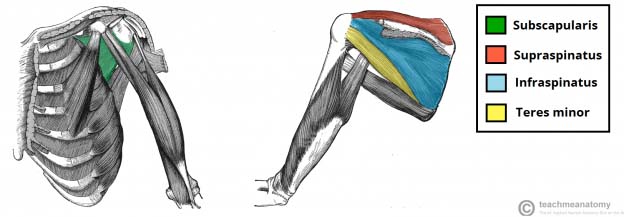
Infraspinatus
…upper limb at the shoulder. Blood supply: Suprascapular and circumflex scapular arteries By TeachMeSeries Ltd (2024) Fig 1 – The rotator cuff muscles, which act to stabilise the shoulder joint….

The Quadrangular Space
…(2024) Fig 1 – Posterior view of the shoulder region, showing the quadrangular space. The subscapularis muscle lies anteriorly, and so cannot be seen on this view. Contents The quadrangular…

Thoracodorsal Artery
…and shoulder. Course The thoracodorsal artery arises on the inferior border of the subscapularis muscle, as a branch of the subscapular artery (itself a branch of the axillary artery). It…

Trapezius
The trapezius is an extrinsic muscle of the shoulder. It is a broad, flat and triangular shape – forming a trapezoid shape in combination with the contralateral side. Attachments: Originates…

Coracobrachialis
…the medial aspect of the humerus shaft (at the level of the deltoid tubercle). Function: Flexion of the arm at the shoulder. It is also a weak adductor at the…

Muscles of the Upper Limb
[child-pages depth=”1″]…

Profunda Brachii Artery (Deep Brachial)
…Supply The profunda brachii artery contributes to the blood supply of the following structures: Shoulder – deltoid Posterior arm – triceps brachii Proxima forearm – anconeus, brachialis, brachioradialis Intermuscular septum…