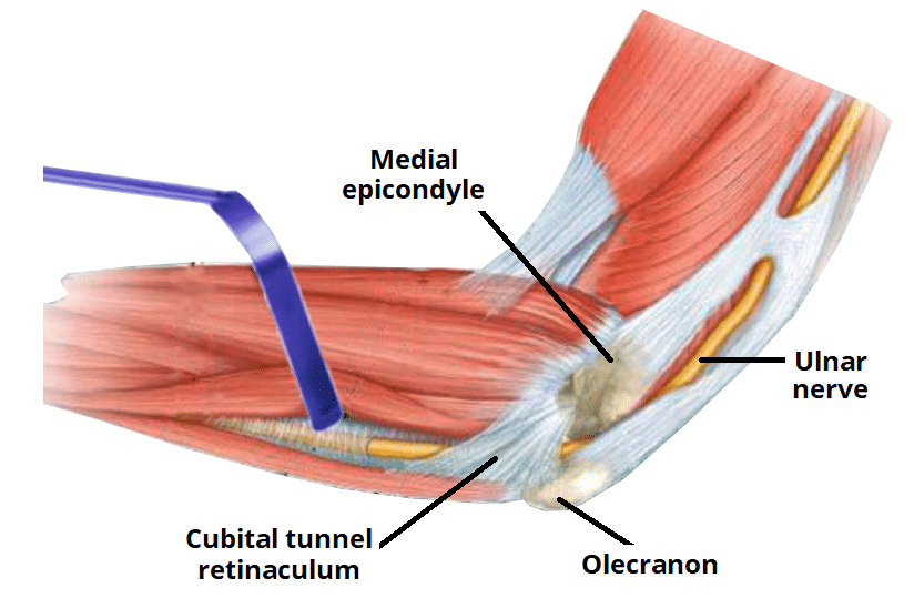
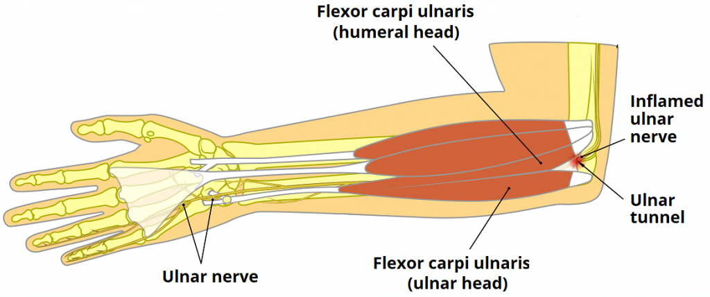
The ulnar (cubital) tunnel is a fibro-osseous space located on the posteromedial aspect of the elbow.
It transmits the ulnar nerve from the arm into the forearm.
In this article, we shall look at the anatomy of the ulnar tunnel – its borders, contents and clinical correlations.
Borders
The ulnar tunnel is oval shaped with medial and lateral walls, a floor, and a roof:
- Medial wall – medial epicondyle of the humerus.
- Lateral wall – olecranon of the ulna.
- Floor – elbow joint capsule and medial collateral ligament of the elbow.
- Roof – ligament spanning between the medial epicondyle and olecranon
The ligament forming the roof of the cubital tunnel is also known as the cubital tunnel retinaculum or the arcuate ligament of Osbourne. It is a band of fascia which runs between the ulnar and humeral heads of the flexor carpi ulnaris.
Contents
The ulnar tunnel transmits the ulnar nerve – a major peripheral nerve of the upper limb. After passing through this space, the ulnar nerve travels between the two heads of the flexor carpi ulnaris and continues into the forearm and hand.
Clinical Relevance: Cubital Tunnel Syndrome
Cubital tunnel syndrome refers to compression of the ulnar nerve within the ulnar tunnel. It is the second most common peripheral neuropathy of the upper limb (after carpel tunnel syndrome).
Patients may experience altered sensation in the sensory distribution of the ulnar nerve – the medial one and half fingers and the associated palm area.
The motor function of the nerve can also be affected in severe disease, causing weakness and wasting of the intrinsic hand muscles (lumbricals, interossei, hypothenar eminence, adductor pollicis).
During elbow flexion, the shape of the cubital tunnel changes from oval to elliptical and the pressure increases. Therefore, this position tends to exacerbate the patient’s symptoms.

