The pineal gland is a small endocrine gland located within the brain. Its main secretion is melatonin, which regulates the circadian rhythm of the body. It is also thought to produce hormones that inhibit the action of other endocrine glands in the body.
In this article, we shall look at the anatomy of the pineal gland – its structure, position and vasculature.
Anatomical Structure and Position
The pineal gland is small glandular body, approximately 6mm long. It is shaped like a pine cone, from which its name is derived. There are two types of cells present within the gland:
- Pinealocytes – hormone secreting cells.
- Glial cells – supporting cells.
In middle age, the gland commonly becomes calcified, and can be subsequently identified on radiographs and CT scans of the head.
Anatomical Position
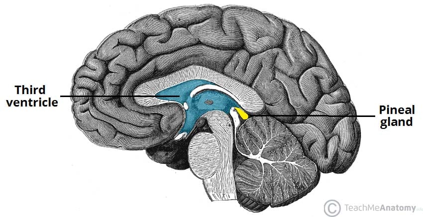
The pineal gland is a midline structure, located between the two cerebral hemispheres. It is attached by a stalk to the posterior wall of third ventricle. In close proximity to the gland are the superior colliculi of the midbrain – paired structures that play an important role in vision.

Fig 1 – Sagittal section of the brain, showing the midline position of the pineal gland
Vasculature
The arterial supply to the pineal gland is profuse, second only to the kidney. The posterior choroidal arteries are the main supply; they are a set of 10 branches that arise from the posterior cerebral artery.
Venous drainage is via the internal cerebral veins.
Clinical Relevance: Pineal Gland Tumours
Pineal glands tumours are a diverse group of neoplasms. The most common is a germ cell tumour, which arises from residual embryonic tissue in the gland.
It presents with the classical symptoms of a space occupying lesion – headache, nausea and vomiting. The tumour can also cause Parinaud syndrome – inability to move the eyes upwards – this is due to compression of the superior colliculi. In addition, obstruction of the cerebral aqueduct may produce hydrocephalus.
In children, a pineal gland tumour (which invades and destroys the gland), produces an accelerated onset of puberty. Thus, it is thought that one of the functions of the gland is to inhibit sexual development.