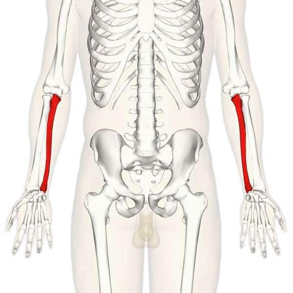
Fig 1.0 – Overview of the anatomical position of the ulna in the upper limb.
The ulna is a long bone in the forearm. It lies medially and parallel to the radius, the second of the forearm bones. The ulna acts as the stabilising bone, with the radius pivoting to produce movement.
Proximally, the ulna articulates with the humerus at the elbow joint. Distally, the ulna articulates with the radius, forming the distal radio-ulnar joint.
In this article, we shall look at the bony landmarks and osteology of the ulna. We shall also consider its clinical correlations.
Proximal Osteology and Articulation
The proximal end of the ulna articulates with the trochlea of the humerus. To enable movement at the elbow joint, the ulna has a specialised structure, with bony prominences for muscle attachment.
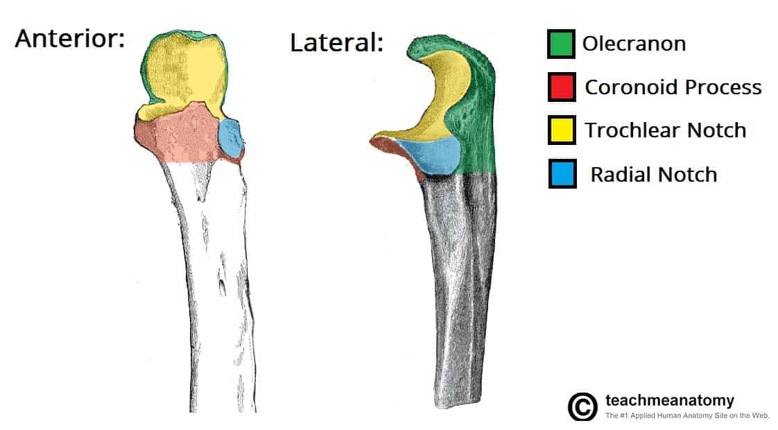
Important landmarks of the proximal ulna are the olecranon, coronoid process, trochlear notch, radial notch and the tuberosity of ulna:
- Olecranon – a large projection of bone that extends proximally, forming part of trochlear notch. It can be palpated as the ‘tip’ of the elbow. The triceps brachii muscle attaches to its superior surface.
- Coronoid process – this ridge of bone projects outwards anteriorly, forming part of the trochlear notch.
- Trochlear notch – formed by the olecranon and coronoid process. It is wrench shaped, and articulates with the trochlea of the humerus.
- Radial notch – located on the lateral surface of the trochlear notch, this area articulates with the head of the radius.
- Tuberosity of ulna – a roughening immediately distal to the coronoid process. It is where the brachialis muscle attaches.
Shaft of the Ulna
The ulnar shaft is triangular in shape, with three borders and three surfaces. As it moves distally, it decreases in width.
The three surfaces:
- Anterior – site of attachment for the pronator quadratus muscle distally.
- Posterior – site of attachment for many muscles.
- Medial – unremarkable.
The three borders:
- Posterior – palpable along the entire length of the forearm posteriorly
- Interosseous – site of attachment for the interosseous membrane, which spans the distance between the two forearm bones.
- Anterior – unremarkable.
Distal Osteology and Articulations
The distal end of the ulna is much smaller in diameter than the proximal end. It is mostly unremarkable, terminating in a rounded head, with distal projection – the ulnar styloid process.
The head articulates with the ulnar notch of the radius to form the distal radio-ulnar joint.
Clinical Relevance – Common Fractures of the Ulna
The forearm is a common site for bone fractures. Here, we shall look at the common fracture types involving the ulna.

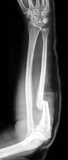
Fig 1.2 – Monteggia fracture of the radius and ulna
A fracture of the ulna alone (not involving the radius) usually occurs as a result of the ulna being hit by an object. The shaft is the most likely site of fracture. In this situation, the normal muscle tone will pull the proximal ulna posteriorly.
Less commonly, the olecranon process can be fractured. This is caused by the patient falling on a flexed elbow. The triceps brachii can displace part of the fragment proximally.
The ulna and the radius are attached by the interosseous membrane. The force of a trauma to one bone can be transmitted to the other via this membrane. Thus, fractures of both the forearm bones are not uncommon.
There are two classical fractures:
- Monteggia’s Fracture – Usually caused by a force from behind the ulna. The proximal shaft of ulna is fractured, and the head of the radius dislocates anteriorly at the elbow.
- Galeazzi’s Fracture – A fracture to the distal radius, with the ulna head dislocating at the distal radio-ulnar joint.
