- The Basics
- Head
- Neuroanatomy
- Neck
- Thorax
- Back
- Upper Limb
- Lower Limb
- Abdomen
- Pelvis
- 3D Body
The Cranial Nerves
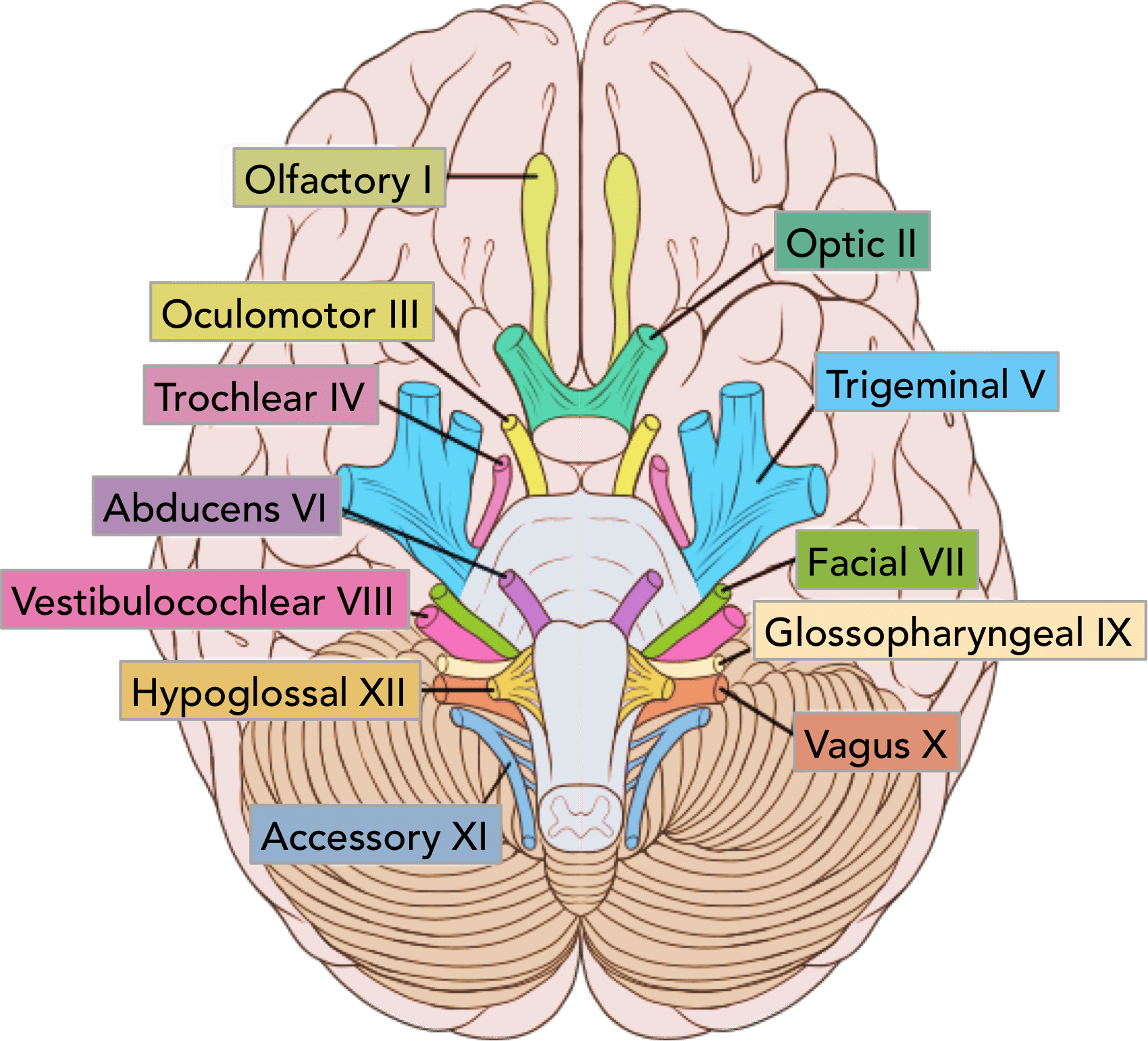
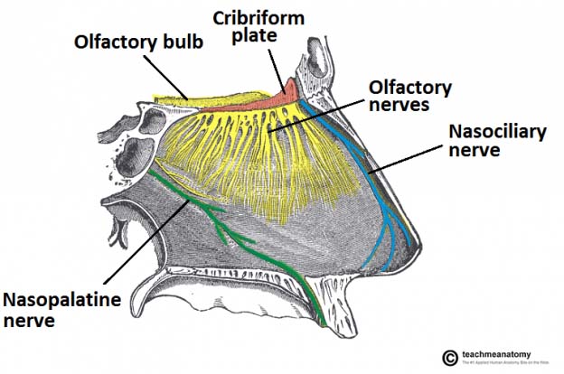
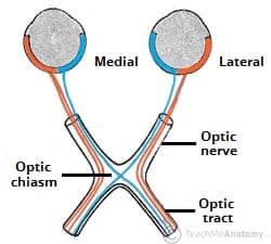
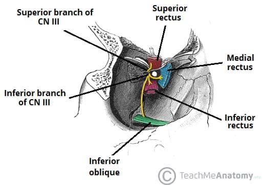
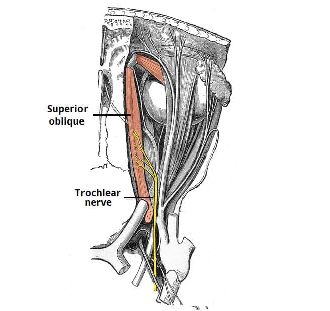
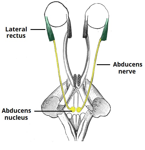
In the section on the cranial nerves, we have articles on each of the 12 cranial nerves. In the first, we discuss the olfactory nerve, detailing its function and describing the anatomy of this important nerve for the sense of smell. The second cranial nerve is the optic nerve, which is responsible for relaying sight back from the retina to the visual cortex in the occipital lobe. Thirdly the oculomotor nerve, which is essential for the movements of the eyeball. It innervates the majority of the extraocular muscles, and along with two other cranial nerves (the trochlear and abducens) it ensures we are able to change our field of vision at will.
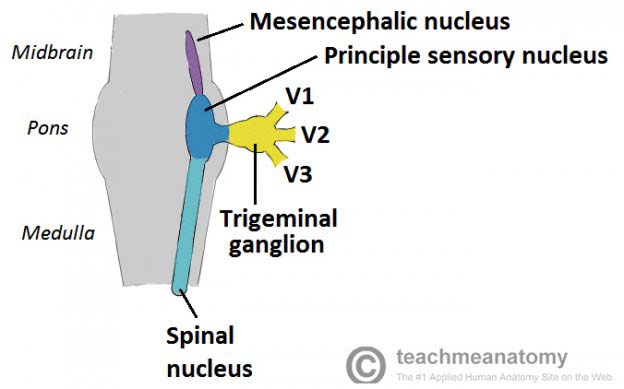
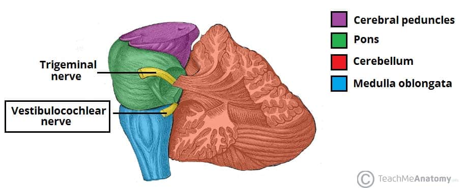
The trigeminal nerve is the fifth paired cranial nerve. It is also the largest cranial nerve. The trigeminal nerve is associated with derivatives of the 1st pharyngeal arch. The three terminal branches of CN V innervate the skin, mucous membranes and sinuses of the face. Their distribution pattern is similar to the dermatome supply of spinal nerves (except there is little overlap in the supply of the divisions). Only the mandibular branch of CN V has motor fibres. It innervates the muscles of mastication: medial pterygoid, lateral pterygoid, masseter and temporalis. Furthermore post-ganglionic neurones of parasympathetic ganglia travel with branches of the trigeminal nerve.
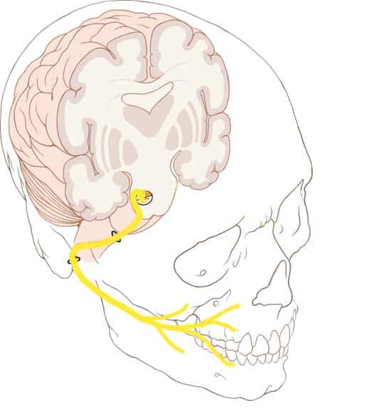
The facial nerve is the seventh paired cranial nerve. In this article, we shall look at the anatomical course of the nerve, and the motor, sensory and parasympathetic functions of its terminal branches. The facial nerve is associated with the derivatives of the second pharyngeal arch. The course of the facial nerve is very complex. There are many branches, which transmit a combination of sensory, motor and parasympathetic fibres.
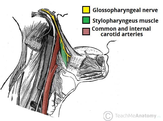
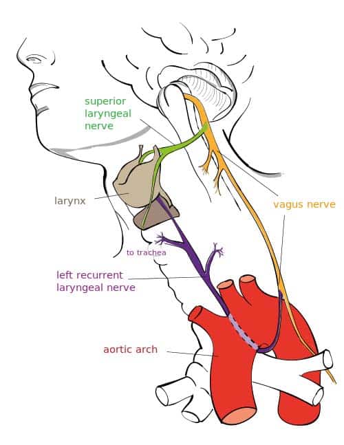
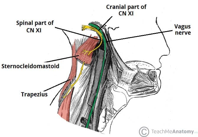
The other cranial nerves are the vestibulocochlear, the glossopharyngeal, the vagus, spinal accessory and hypoglossal nerves. They are all discussed in great detail in their respective articles.
