The triangular interval is an anatomical space located immediately below the axilla region.
It allows structures to pass between the anterior and posterior compartments of the upper arm, as well as between the posterior compartment and the axilla.
In this article, we shall look at the anatomy of the triangular interval – its borders, contents and clinical correlations.
Borders
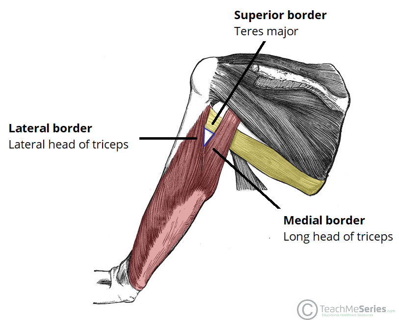
The triangular interval is orientated with the base superiorly and apex inferiorly. It has three borders:
- Superior – Inferior border of the teres major.
- Lateral – Shaft of the humerus and lateral head of the triceps brachii.
- Medial – Lateral border of the long head of the triceps brachii.
Contents
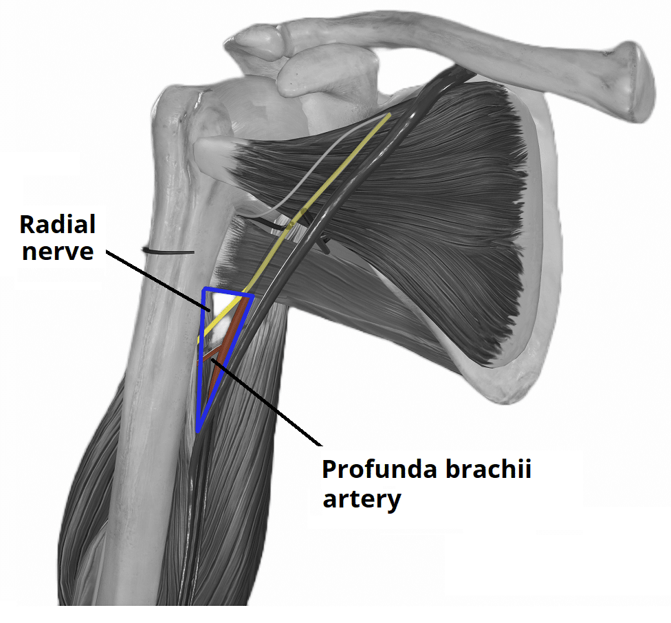
The triangular interval is a passageway that allows structures to travel between:
- Anterior and posterior compartments of the upper arm
- Posterior compartment of the upper arm and the axilla
It contains the radial nerve and profunda brachii artery (and accompanying vena comitantes), as they travel into the posterior compartment of the upper arm.
Clinical Relevance: Triangular Interval Syndrome
Triangular interval syndrome refers to compression of the radial nerve as it passes through the triangular interval.
It is thought to occur secondary to hypertrophy of triceps brachii or teres major. The nerve may also be compressed by fibrous bands spanning between these two muscles.
Clinical features of triangular interval syndrome include:
- Neuropathic pain or paraesthesia in the sensory distribution of the radial nerve.
- Weakness in extension of the elbow, wrist, or digits.

