- The Basics
- Head
- Neuroanatomy
- Neck
- Thorax
- Back
- Upper Limb
- Lower Limb
- Abdomen
- Pelvis
- 3D Body
Areas of the Head
The areas of the head include: the scalp, the infratemporal fossa, the pterygopalatine fossa, and the cranial fossae.
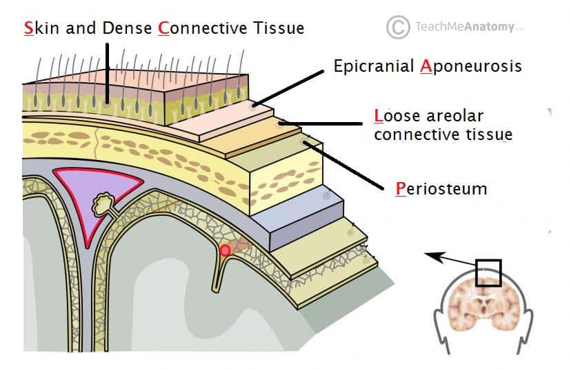
Overlying the cranial bones, the scalp consists of 5 layers: skin, connective tissue (dense), aponeurosis, loose connective tissue and the periosteum. The three outermost layers move as one unit, with the aponeurosis being a tendon-like structure spanning between the frontalis and occipitalis muscles. The scalp receives a rich arterial supply from the external carotid artery, and sensory innervation from the trigeminal nerve, as well as cervical nerves.
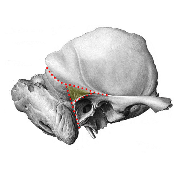
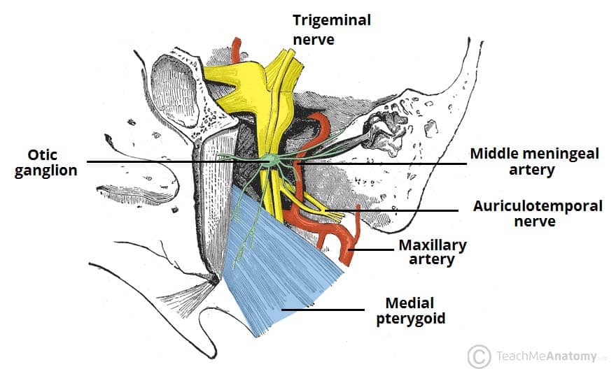
The infratemporal fossa is a complex area located at the base of the skull, deep to the masseter muscles. The infratemporal fossa provides a conduit for neurovascular structures entering and leaving the cranial cavity. The key neurovascular structures located in the infratemporal fossa are the mandibular nerve, chorda tympani and otic ganglion, the maxillary artery, pterygoid venous plexus, maxillary vein, and middle meningeal vein. The lateral and medial pterygoid muscles of mastication are also located in the infratemporal fossa.
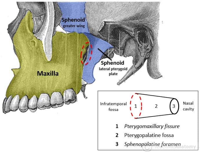
Located deep to the infratemporal fossa, the pterygopalatine fossa extends to the nasal cavity. The key contents of the pterygopalatine fossa include: the maxillary nerve and its branches, the pterygopalatine ganglion, as well as the maxillary artery and its branches.
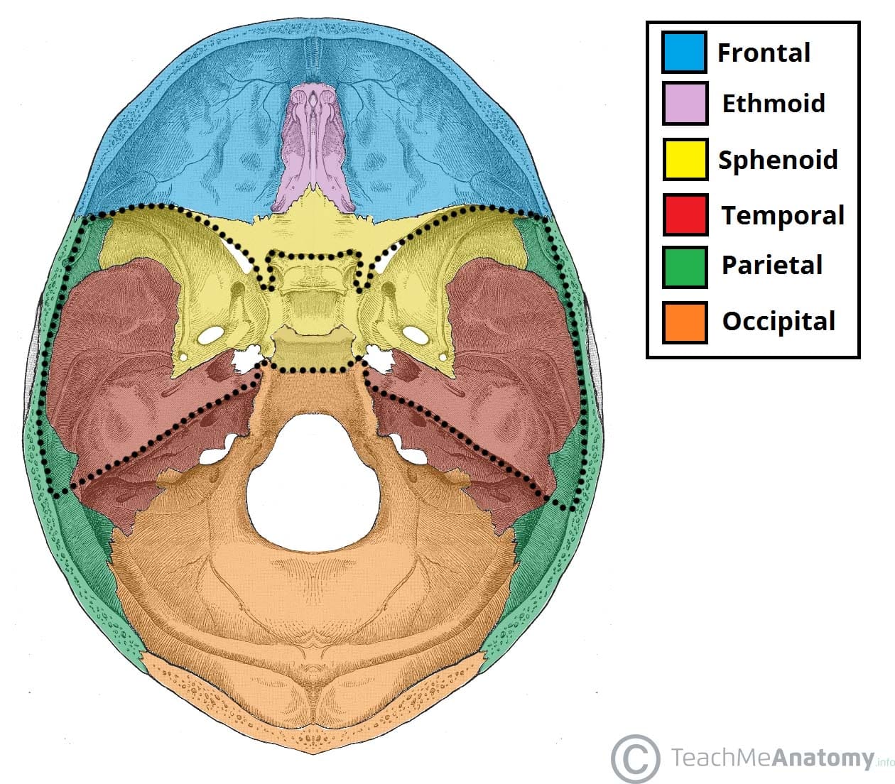
The floor of the cranial cavity can be split in to three key areas: the anterior, middle and posterior cranial fossae. Each fossa accommodates and supports different parts of the brain, as well as allowing passage of neurovasculature in and out of the cranial cavity. The anterior cranial fossa accommodates the frontal lobes, the olfactory bulb, and the anterior and posterior ethmoidal neurovascular structures. The middle cranial fossa accommodates the pituitary glands, temporal lobes, and the optic canals. Neurovascular structures pass through the supraorbital fissure, foramen rotundum, foramen ovale and foramen spinosum, all of which are accommodated by the middle cranial fossa. The posterior cranial fossa accommodates the brainstem and the cerebellum.
In this section, learn more about the areas of the head including the scalp, the infratemporal fossa, the pterygopalatine fossa, and the cranial fossae.
