The flexor pulley system of the hand is a complex structure that co-ordinates flexion of the digits. It consists of:
- Long flexor tendons – and their associated synovial sheaths.
- Annular pulleys – 5 associated with each finger, 2 associated with the thumb.
- Cruciate pulleys – 3 associated with each finger.
- Oblique pulley – 1 associated with the thumb.
The main role of the flexor pulley system is to hold the flexor tendons against the phalanges; preventing them from pulling away and bowstringing. This allows efficient flexion of the individual digits by the long flexor muscles.
In this article, we shall look at the anatomy of the flexor pulley system – including the fibrous digital sheaths, annular ligaments and cruciate ligaments.
Flexor Pulley System of the Fingers
Long Flexor Tendons
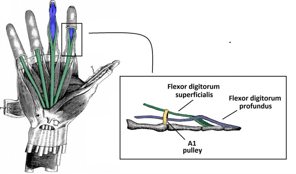
The long flexor tendons of the fingers arise from the flexor digitorum superficialis (FDS) and flexor digitorum profundus (FDP) forearm muscles. These tendons enter the hand via the carpal tunnel, enclosed in a common synovial sheath.
Within the hand, the tendons fan out and enter their respective fibrous flexor sheaths. These sheaths are strong ligamentous tunnels, each associated with a digit.
At the base of the proximal phalanx, the FDS tendon splits into two – with the FDP tendon passing between them. The split FDS tendons attach to the base of the middle phalanx, whilst the FDP tendon attaches to the base of the distal phalanx.
Annular and Cruciate Pulleys
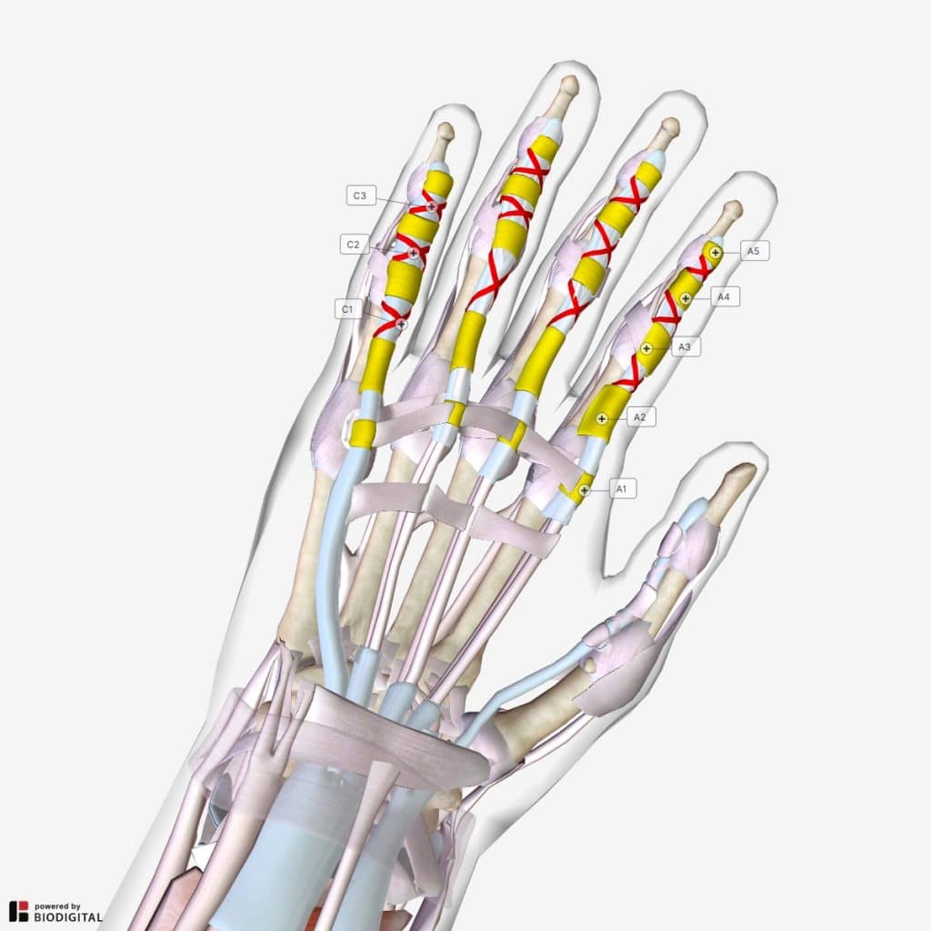
The fibrous flexor sheaths contain thickened areas – known as the annular and cruciform pulleys.
The annular pulleys (or ligaments) represent five areas where the fibrous flexor sheaths are reinforced by circular fibres. It is thought that A2 and A4 are most important in preventing bowstringing of the flexor tendons.
- A1 – overlies the metacarpophalangeal joint
- A2 – overlies the proximal aspect of the proximal phalanx
- A3 – overlies the proximal interphalangeal joint
- A4 – overlies the mid-portion of the middle phalanx
- A5 – overlies the distal interphalangeal joint
The cruciate pulleys refer to areas where the fibrous flexor sheaths are reinforced by cruciform fibres:
- C1 – located between A2 and A3
- C2 – located between A3 and A4
- C3 – located between A4 and A5
Flexor Pulley System of the Thumb
The long flexor tendon of the thumb arises from the flexor pollicis longus (FPL). It passes into the hand through the carpal tunnel.
The FPL tendon then enters the fibrous flexor sheath of the thumb and attaches to the base of the distal phalanx.
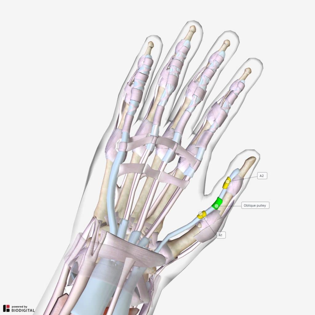
The fibrous flexor sheath of the thumb is reinforced by three pulleys:
- A1 – overlies the metacarpophalangeal joint
- Oblique – overlies the proximal half of proximal phalanx
- A2 – overlies the distal half of proximal phalanx

Fig 3 – The flexor pulley system of the thumb is formed by the A1, oblique and A2 pulleys
Clinical Relevance: Trigger Finger
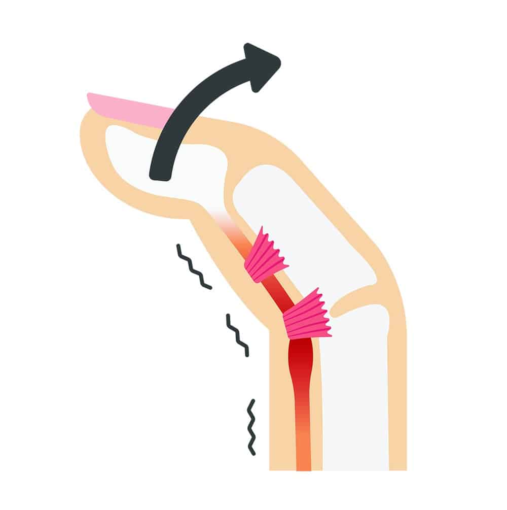
Trigger finger is a condition in which the finger or thumb click or lock when in flexion, preventing a return to extension.
It is thought to be caused by inflammation of the long flexor tendons (often from repetitive movements of the fingers). In response to the inflammation, the tendon becomes thickened or develops a nodule – making it difficult for the tendon to pass through the pulleys. The A1 pulley is the most frequently involved ligament in trigger finger.
When the fingers are flexed, the node moves proximal to the pulley, however when the patient attempts to extend the digit this node fails to pass back under the pulley. Consequently, the digit becomes locked in a flexed position.
Management options including splinting of the affecting digit, steroid injections and surgical release of the tendon.


