The mediastinum is the central compartment of the thoracic cavity, located between the two pleural sacs. It contains most of the thoracic organs, and acts as a conduit for structures traversing the thorax on their way into the abdomen.
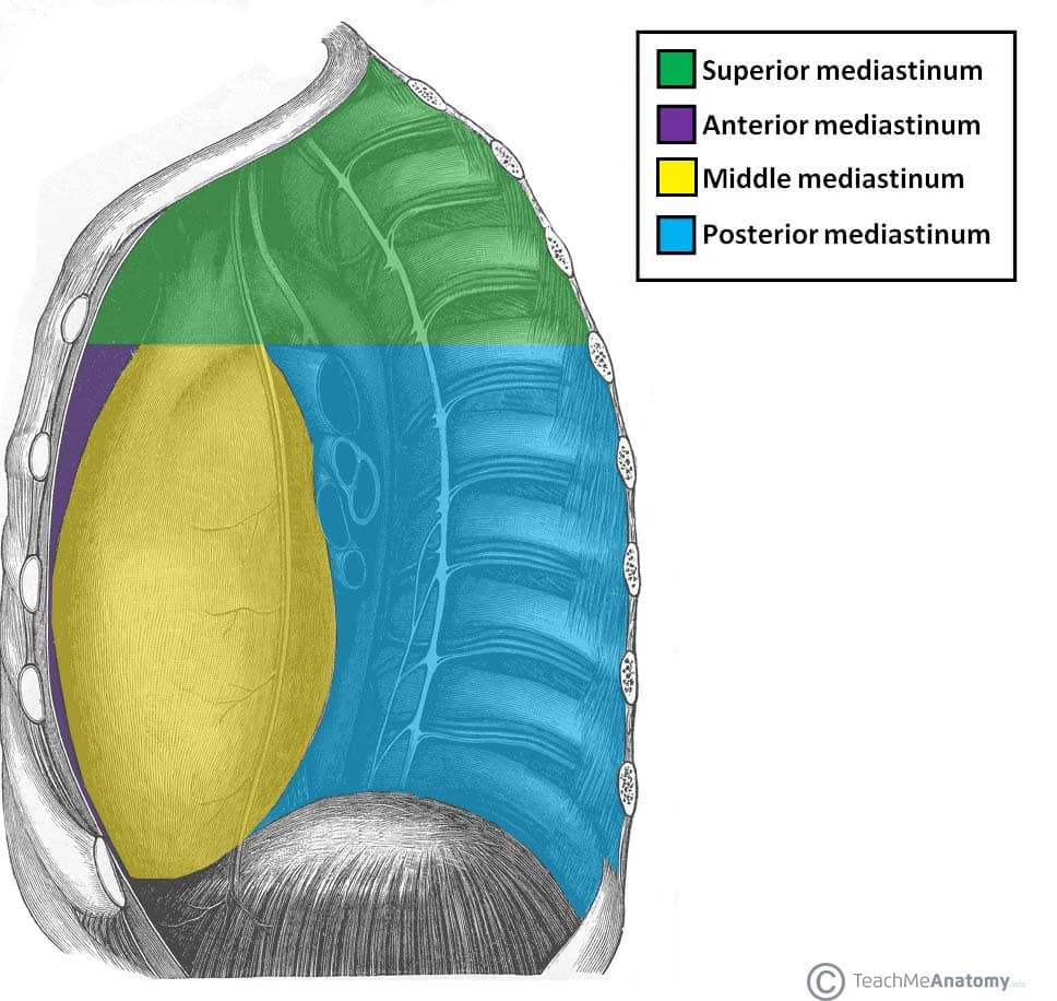
Anatomically, the mediastinum is divided into two parts by an imaginary line that runs from the sternal angle (the angle formed by the junction of the sternal body and manubrium) to the T4 vertebrae:
- Superior mediastinum – extends upwards, terminating at the superior thoracic aperture.
- Inferior mediastinum – extends downwards, terminating at the diaphragm. It is further subdivided into the anterior mediastinum, middle mediastinum and posterior mediastinum.
In this article, we shall look at the anatomy of the anterior mediastinum – its borders, contents and clinical correlations.
Borders
The anterior mediastinum is bordered by the following thoracic structures:
- Lateral borders: Mediastinal pleura (part of the parietal pleural membrane).
- Anterior border: Body of the sternum and the transversus thoracis muscles.
- Posterior border: Pericardium.
- Roof: Continuous with the superior mediastinum at the level of the sternal angle.
- Floor: Diaphragm.
Contents
The anterior mediastinum contains no major structures. It accommodates loose connective tissue (including the sternopericardial ligaments, which tether the pericardium to the sternum), fat, some lymphatic vessels, lymph nodes and branches of the internal thoracic vessels.
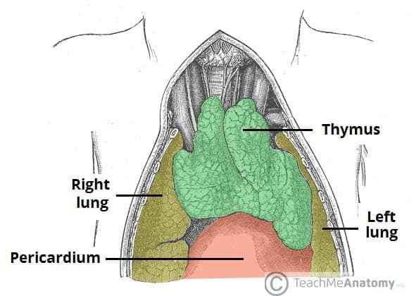
In infants and children, the thymus extends inferiorly into the anterior mediastinum. However the thymus recedes during puberty and is mostly replaced by adipose tissue in the adult.

Fig 1.1 – The anatomical position of the thymus. It is mostly located in the superior mediastinum, but can extend into the anterior mediastinum.
