The adductor canal (Hunter’s canal, subsartorial canal) is a narrow conical tunnel located in the thigh.
It is approximately 15cm long, extending from the apex of the femoral triangle to the adductor hiatus of the adductor magnus. The canal serves as a passageway from structures moving between the anterior thigh and posterior leg.
In this article, we shall look at the anatomy of the adductor canal – its borders, contents and clinical relevance.
Borders
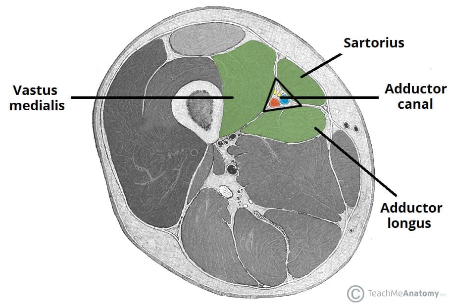
The adductor canal is bordered by muscular structures:
- Anteromedial – Sartorius.
- Lateral – Vastus medialis.
- Posterior – Adductor longus and adductor magnus.
The adductor canal runs from the apex of the femoral triangle to the adductor hiatus – a gap between the adductor and hamstring attachments of the adductor magnus muscle.

Fig 1 – Cross-section of the thigh, showing the borders of the adductor canal. Note: the adductor magnus is not visible in this illustration.
Contents
The adductor canal serves as a passageway for structures moving between the anterior thigh and posterior leg.
It transmits the femoral artery, femoral vein (posterior to the artery), nerve to the vastus medialis and the saphenous nerve – the largest cutaneous branch of the femoral nerve.
As the femoral artery and vein exit the canal, they are called the popliteal artery and vein respectively.
Clinical Relevance – Adductor Canal Block
In the adductor canal block, local anaesthetic is administered in the adductor canal to block the saphenous nerve in isolation, or together with the nerve to the vastus medialis.
The block can be used to provide sensory anaesthesia for procedures involving the distal thigh and femur, knee and lower leg on the medial side. The sartorius and femoral artery are used as anatomical landmarks to locate the saphenous nerve.
Clinical Relevance – Adductor Canal Compression Syndrome
Adductor canal compression syndrome describes entrapment of the neurovascular bundle within the adductor canal. A rare condition, it is usually caused by hypertrophy of adjacent muscles such as vastus medialis.
It is most common in young males, who may present with claudication symptoms due to femoral artery occlusion (more common) or neurological symptoms due to entrapment of the saphenous nerve.