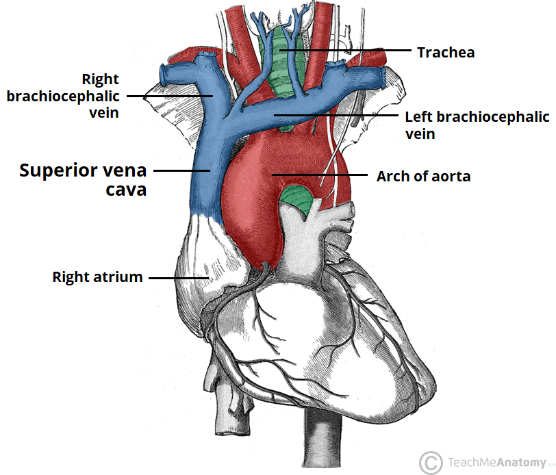
The superior vena cava is formed from the unification of the left and right brachiocephalic veins and
carries blood from the top half of the body and into the right atrium. Unlike most veins, the superior vena cava does not have any valves, and this is important to remember clinically when wanting to determine the pressure in the right atrium.
In addition to the superior vena cava, the vasculature of the thoraxalso includes the aorta. The aorta is the largest artery in the body and acts as a conduit for blood to supply the rest of the body via the systemic circulation. The aorta consists of 4 major parts: the ascending aorta, the aortic arch, the thoracic aorta and the abdominal aorta. The ascending aorta contains the left and right aortic sinuses, which is where the left and right coronary arteries will originate. The arch of the aorta contains 3 major branches: the brachiocephalic trunk, the left common carotid artery and the left subclavian artery. The thoracic aorta descends in the thoracic cavity and pierces the diaphragm where it will then continue as the abdominal aorta. There are many branches that arise from both the thoracic and abdominal aorta and are important for supplying local organs with oxygenated blood. When the abdominal aorta reaches the level of the L4 vertebra, it bifurcates into the right and left common iliac arteries which go on to supply the lower body.
In this section, learn more about the vasculature of the thorax– the superior vena cava and the aorta.


