The venous system of the upper limb drains deoxygenated blood from the arm, forearm and hand. It can be subdivided into the superficial system and the deep system.
In this article, we shall look at the anatomy of the upper limb veins – their anatomical course, structure, and their clinical relevance.
Superficial Veins
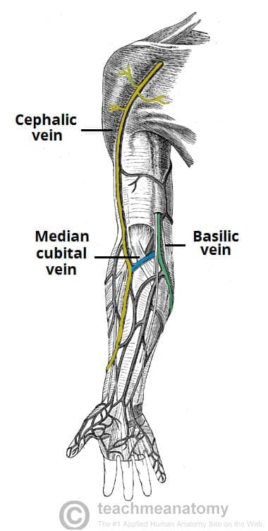
The major superficial veins of the upper limb are the cephalic and basilic veins. They are located within the subcutaneous tissue of the upper limb.
Basilic Vein
The basilic vein originates from the dorsal venous network of the hand and ascends the medial aspect of the upper limb.
At the border of the teres major, the vein moves deep into the arm. Here, it combines with the brachial veins from the deep venous system to form the axillary vein.
Cephalic Vein
The cephalic vein also arises from the dorsal venous network of the hand. It ascends the antero-lateral aspect of the upper limb, passing anteriorly at the elbow.
At the shoulder, the cephalic vein travels between the deltoid and pectoralis major muscles (known as the deltopectoral groove), and enters the axilla region via the clavipectoral triangle. Within the axilla, the cephalic vein empties into axillary vein.
The cephalic and basilic veins are connected at the elbow by the median cubital vein.
Deep Veins
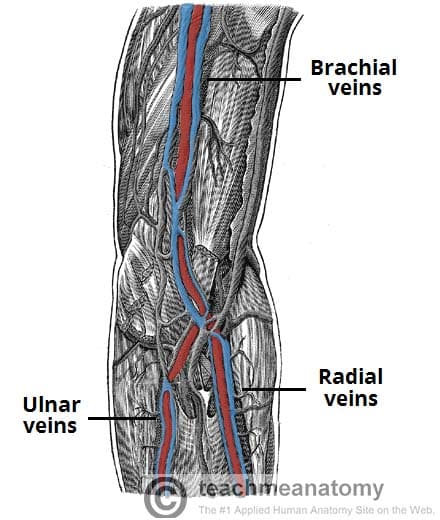
The deep venous system of the upper limb is situated underneath the deep fascia. It is formed by paired veins, which accompany and lie either side of an artery. In the upper extremity, the deep veins share the name of the artery they accompany.
The brachial veins are the largest in size, and are situated either side of the brachial artery. The pulsations of the brachial artery assist the venous return. Veins that are structured in this way are known as vena comitantes.
Perforating veins run between the deep and superficial veins of the upper limb, connecting the two systems.
Clinical Relevance: Venepuncture
Venepuncture is the practice of obtaining intravenous access. This is usually for the purpose of providing intravenous therapy (e.g. fluids, medications) or for obtaining a blood sample.
The median cubital vein is a common site of venepuncture. It is a superficial vein that is located anteriorly to the cubital fossa region. It is thought to be fixed in place by perforating veins, which arise from the deep venous system and pierce the bicipital aponeurosis.
Its ease of access, fixed position and superficial position make the median cubital vein a good site for venepuncture in many individuals.

