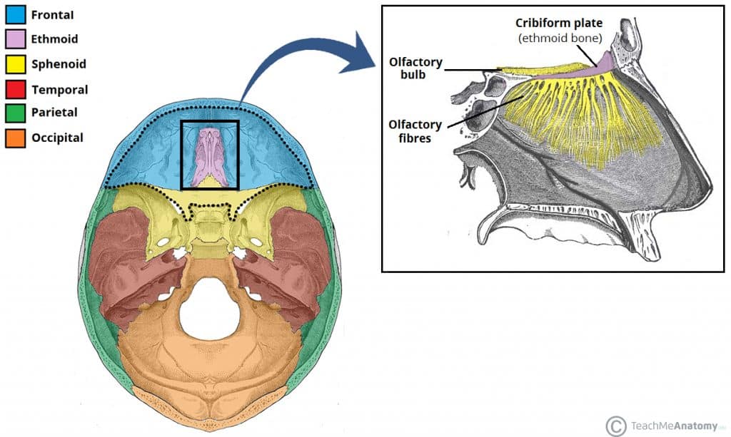
The ethmoid bone is a small unpaired bone, located in the midline of the anterior cranium – the superior aspect of the skull that encloses and protects the brain.
The term ‘ethmoid’ originates from the Greek ‘ethmos’, meaning sieve. This is reflected in its lightweight, spongy structure.
In this article, we shall look at the anatomy of the ethmoid bone – its location, relations, and structure.
Anatomical Structure
The ethmoid bone is one of the 8 bones of the cranium. It is situated at the roof of the nasal cavity, and between the two orbital cavities.
It contributes to the medial wall of the orbit and forms part of the anterior cranial fossa, where it separates the nasal cavity (inferiorly) from the cranial cavity (superiorly). It also forms a significant portion of the nasal septum and lateral nasal wall.
The olfactory nerve (CN I) has a close anatomical relationship with the ethmoid bone. Its numerous nerve fibres pass through the cribriform plate of the ethmoid bone to innervate the nasal cavity with the sense of smell.
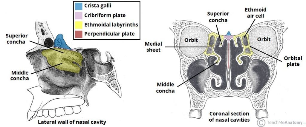
The ethmoid bone is made up of three parts – the cribriform plate, the perpendicular plate, and the ethmoidal labyrinth.
The cribriform plate forms the roof of the nasal cavity. It is pierced by numerous olfactory nerve fibres, which gives it a sieve-like structure. Projecting superiorly from the cribriform plate is the crista galli, which provides an attachment point for the falx cerebri (sheet of dura mater that separates the two cerebral hemispheres).
Another projection of bone descends from the cribriform plate – the perpendicular plate. It forms the superior two-thirds of the nasal septum.
Lastly, the ethmoid bone contains two ethmoidal labyrinths. These are large masses located at either side of the perpendicular plate, which contain the ethmoidal air cells (sinuses). Two sheets of bone form each labyrinth:
- Orbital plate – the lateral sheet of bone, which also forms the medial wall of the orbit
- Medial sheet – forms the upper lateral wall of the nasal cavity, from which the superior and middle conchae extend into the nasal cavity.
Articulations
The ethmoid bone articulates with 13 others:
- Paired – nasal bones, maxillae, lacrimal bones, palatine bones, inferior conchae.
- Unpaired – frontal, vomer and sphenoid bones.
Clinical Relevance – Ethmoid Fracture
The ethmoid bone can be fractured in cases of facial trauma – most commonly hitting the dashboard in a collision, or a fall from height. Some signs and symptoms of fracture are related to the anatomy of the ethmoid bone:
- Fracture of cribriform plate – branches of the olfactory bulb may be sheared. This may cause anosmia (loss of sense of smell).
- Fracture of the labyrinth – may allow communication between the nasal cavity and the orbit. It is then possible for air to enter the orbit and cause orbital emphysema.
Clinical Relevance – CSF Rhinorrhoea
A fracture to the cribriform plate may allow communication between the nasal cavity and the central nervous system. Consequently, cerebrospinal fluid (CSF) can enter the nasal cavity and drain out from the nose. This manifests clinically as a clear watery discharge from one side of the nose – and is known as CSF rhinorrhoea.
The leaks normally stop spontaneously and can be managed conservatively, however surgery is sometimes required. Spontaneous CSF rhinorrhoea can also occur due to congenital or acquired defects in the ethmoid bone.

