Fascia is a sheet or band of fibrous tissue lying deep to the skin. It lines, invests, and separates structures within the body. There are three main types of fascia:
- Superficial fascia – blends with the reticular layer beneath the dermis.
- Deep fascia – envelopes muscles, bones, and neurovascular structures.
- Visceral fascia – provides membranous investments that suspend organs within their cavities.
In this article, we shall look at the anatomy of the deep fascia of the thigh – the fascia lata. A detailed account of its structure, anatomical relations, function, attachments, and clinical relevance will be outlined.
Anatomical Structure
The fascia lata is a deep fascial investment of the musculature of the thigh, and is analogous to a strong, extensible, and elasticated stocking. It begins proximally around the iliac crest and inguinal ligament and ends distal to the bony prominences of the tibia. It is continuous with what is renamed the deep fascia of the leg (also known as the crural fascia).
The depth of the fascia lata varies considerably across the thigh. It is thickest along the superolateral aspect of the thigh, where it arises from the fascial condensations of gluteus maximus and medius. It is also thick around the knee where the fascia receives reinforcing fibres from tendons of the quadriceps muscles. The fascial investment is thinnest where it covers the adductor muscles of the medial thigh.
The deepest aspect of the fascia lata gives rise to three intermuscular septa that attach centrally to the femur:
- Medial – separates the anterior thigh compartment from the medial compartment.
- Lateral – separates the anterior thigh compartment from the posterior compartment.
- Posterior – separates the medial thigh compartment from the posterior compartment.
The lateral intermuscular septum is the strongest of the three due to reinforcement from the iliotibial tract (see later), whereas the other two are proportionately weaker.
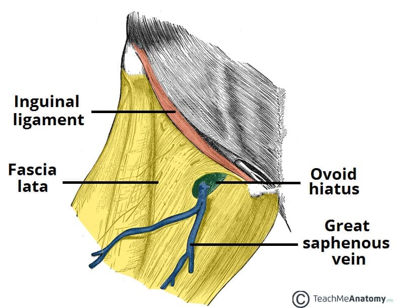
An ovoid hiatus known as the saphenous opening is present in the fascia lata just inferior to the inguinal ligament. The opening serves as an entry point for efferent lymphatic vessels and the great saphenous vein, draining into superficial inguinal lymph nodes and the femoral vein respectively.
Clinical Significance: Femoral Hernia
A femoral hernia develops when an out-pouching of abdominal viscera protrudes through the femoral canal. The protrusion becomes noticeable when it exits superficially through the saphenous opening within the fascia lata – producing a swelling inferior to the inguinal ligament.
The saphenous opening is relatively small, and the surrounding fascia lata is quite tight and inflexible, so femoral hernias carry high risk of bowel incarceration or strangulation, which requires rapid surgical intervention.
Anatomical Relations
Iliotibial Tract (ITT)
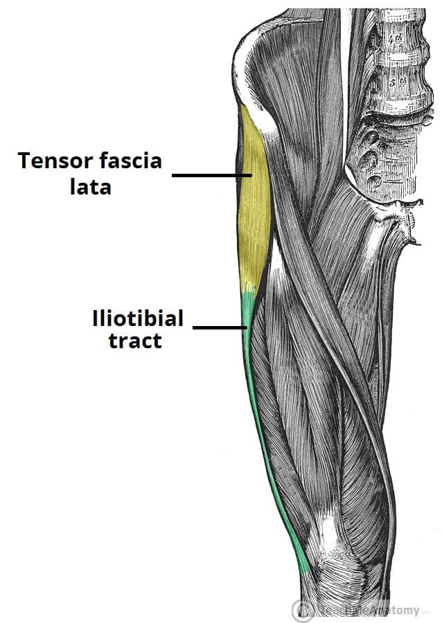
The iliotibial tract (sometimes known as the iliotibial band or IT band) is a longitudinal thickening of the fascia lata, which is strengthened superoposteriorly by fibres from the gluteus maximus.
It is located laterally in the thigh, extending from the iliac tubercle to the lateral tibial condyle. The ITT has three main functions:
- Movement – acts as an extensor, abductor and lateral rotator of the hip, with an additional role in providing lateral stabilisation to the knee joint.
- Compartmentalisation – the deepest aspect of ITT extends centrally to form the lateral intermuscular septum of the thigh and attaches to the femur.
- Muscular sheath – forms a sheath around the tensor fascia lata muscle.
Tensor Fascia Lata (TFL)
The tensor fascia lata is a gluteal muscle that acts as a flexor, abductor, and internal rotator of the hip. Its name, however, is derived from its additional role in tensing the fascia lata. It is innervated by the superior gluteal nerve, like gluteus medius and minimus, but is located more anterolaterally than the other gluteal muscles.
The muscle originates from the iliac crest and descends inferiorly to the superolateral thigh. At the junction of the middle and upper thirds of the thigh, it inserts into the anterior aspect of the iliotibial tract. When stimulated, the tensor fasciae lata tautens the iliotibial band and braces the knee, especially when the opposite foot is lifted.
The property of TFL tightening the fascia lata is analogous to hoisting an elastic stocking up the thigh. When the fascia lata is pulled taut, it forces muscles in the anterior and posterior compartments closer to towards the femur. Contraction within each compartment centralises muscle weight and limits outward expansion, which in turn reduces the overall force required for movement at the hip joint.
An additional property of tensing the fascia lata is that it makes muscle contraction more efficient in compressing deep veins, which ensures adequate venous return to the heart from the lower limbs.
Attachments
Proximal
The fascia lata arises from multiple superior attachments around the pelvis and hip region:
- Posterior – sacrum and coccyx.
- Lateral – iliac crest.
- Anterior – inguinal ligament, superior pubic rami.
- Medial – inferior ischiopubic rami, ischial tuberosity, sacrotuberous ligament.
The fascia lata is also continuous with other regions of deep and superficial fascia at its superior aspect. The deep iliac fascia descends from the thoracic region at the diaphragm, covers the entire iliacus and psoas regions, and blends with the fascia lata superiorly.
The deep layer of the superficial fascia of the abdominal wall (Scarpa’s fascia) blends with the fascia lata just below the inguinal ligament.
Lateral
The lateral thickening of fascia lata forms the iliotibial tract and receives tendon insertions superiorly from gluteus maximus and tensor fascia lata. The widened band of fibres descends the lateral thigh and attaches to the lateral tibial condyle on the anterolateral (Gerdy) tubercle.
Inferior
The fascia lata ends at the knee joint where it then becomes the deep fascia of the leg (the crural fascia). Attachments are made at bony prominences around the knee including the femoral and tibial condyles, patella, head of fibula and the tibial tuberosity.
Central
The deep aspect of fascia lata produces three intermuscular septa which attach centrally to the femur. The lateral septum joins to the lateral lip of the linea aspera and the medial and anterior septa attach to the medial lip. These attachments then continue along the whole length of the femur to include the supracondylar lines.
Clinical Significance: Transplantation
Dermatofasciotomy and debridement can leave large wound sites that require post-operative grafts to facilitate tissue regeneration and healing. The fascia lata graft is a popular choice as the iliotibial tract provides a particularly high concentration of connective tissue fibres, and can be surgically harvested whilst leaving the majority of fibres intact.
Novel developments in transplantation have also shown success with using fascia lata in reconstructive surgery. Advances have included:
- Heart valve replacements
- Eyelid reparations
- Dura mater repair
- Urinary incontinence treatment (fascia lata sling)
The main advantage of using fascia lata as opposed to an artificial product is that the native tissue is well vascularised upon transplantation, whereas the latter may require microvascular anastomosis.

