The veins of the lower limb drain deoxygenated blood and return it to the heart. They can be divided into two groups – deep and superficial:
- Deep veins are located underneath the deep fascia of the lower limb, accompanying the major arteries.
- Superficial veins are found in the subcutaneous tissue. They eventually drain into the deep veins.
In this article, we shall examine the anatomy and clinical correlations of the major veins of the lower limb.
Deep Veins of the Lower Limb
The deep venous drainage system of the lower limb is located beneath the deep fascia of the lower limb.
As a general rule, the deep veins accompany and share the name of the major arteries in the lower limb. Often, the artery and vein are located within the same vascular sheath – so that the arterial pulsations aid the venous return.
Foot and Leg
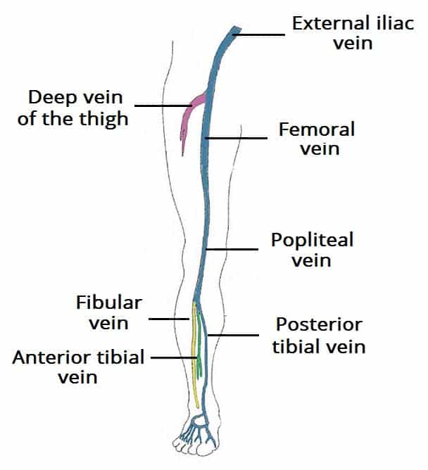
The main venous structure of the foot is the dorsal venous arch, which mostly drains into the superficial veins. Some veins from the arch penetrate deep into the leg, forming the anterior tibial vein.
On the plantar aspect of the foot, medial and lateral plantar veins arise. These veins combine to form the posterior tibial and fibular veins. The posterior tibial vein accompanies the posterior tibial artery, entering the leg posteriorly to the medial malleolus.
On the posterior surface of the knee, the anterior tibial, posterior tibial and fibular veins unite to form the popliteal vein. The popliteal vein enters the thigh via the adductor canal.
Thigh
Once the popliteal vein has entered the thigh, it is known as the femoral vein. It is situated anteriorly, accompanying the femoral artery.
The deep vein of the thigh (profunda femoris vein) is the other main venous structure in the thigh. Via perforating veins, it drains blood from the thigh muscles. It then empties into the distal section of the femoral vein.
The femoral vein leaves the thigh by running underneath the inguinal ligament, at which point it is known as the external iliac vein.
Gluteal Region
The gluteal region is drained by inferior and superior gluteal veins. These empty into the internal iliac vein.
Clinical Relevance: Deep Vein Thrombosis
Deep vein thrombosis (DVT) is the formation of the blood clot within the deep veins of the lower limbs, causing blockage of the vessel. Locally, this causes pain, swelling and tenderness of the affected limb.
The main complication of a DVT is pulmonary embolism. The thrombus can become dislodged, and travel into pulmonary circulation. Pulmonary occlusion prevents blood from returning to the heart, resulting in mechanical shock.
Patients that are considered high risk of developing a DVT undergo prophylactic treatment to prevent thrombosis.
Superficial Veins of the Lower Limb
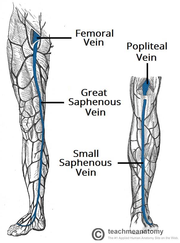
The superficial veins of the lower limb run in the subcutaneous tissue. There are two major superficial veins – the great saphenous vein, and the small saphenous vein.
Long Saphenous Vein
The long saphenous vein is formed by the dorsal venous arch of the foot, and the dorsal vein of the great toe. It ascends up the medial side of the leg, passing anteriorly to the medial malleolus at the ankle, and posteriorly to the medial condyle at the knee.
As the vein moves up the leg, it receives tributaries from other small superficial veins. The great saphenous vein terminates by draining into the femoral vein immediately inferior to the inguinal ligament.
Surgically, the great saphenous vein can be harvested and used as a vessel in coronary artery bypasses.
Small Saphenous Vein
The small saphenous vein is formed by the dorsal venous arch of the foot, and the dorsal vein of the little toe. It moves up the posterior side of the leg, passing posteriorly to the lateral malleolus, along the lateral border of the calcaneal tendon.
At the level of the knee, the short saphenous vein passes between the two heads of the gastrocnemius muscle and empties into the popliteal vein in the popliteal fossa.
Clinical Relevance: Varicose Veins
In the lower limbs, venous blood flows from the skin to superficial veins, which drain into the deep veins.
With the veins there are valves that prevent back flow of blood. If these valves become incompetent, blood can flow back into the superficial veins. This results in an increased intra-luminal pressure, which the veins cannot withstand, causing them to become dilated and tortuous. This condition is known as varicose veins.
There are various soft tissue changes that can occur with chronic varicose veins. Due to the incompetence of the valves, the pressure in the venous system rises. This damages the cells, causing blood to extrude into skin. Further complications can produce a brown pigmentation and ulceration of the surrounding tissue.
Varicose veins can be treated by;
-
- Vein ligation, stripping, and avulsion – making an incision in the groin (or popliteal fossa) and identifying the responsible vein, before tying it off and stripping it away.
- Foam sclerotherapy – injecting a sclerosing (irritating) agent directly into the varicosed veins, causing an inflammatory response that closes off the vein.
- Thermal ablation – heating the vein from inside (via radiofrequency or laser catheters), causing irreversible damage to the vein which closes it off.
By Jackerhack [CC-BY-SA-2.5], from Wikimedia Commons
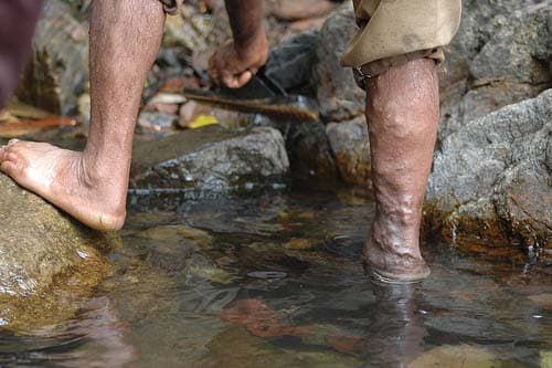
Fig 3 – Varicose veins on the right leg.