The profunda femoris (deep femoral) artery is the largest branch of the femoral artery.
It arises within the anterior thigh, and travels deeply to supply the muscles and skin of the lateral, medial and posterior thigh.
Pro Feature - 3D Model
Course
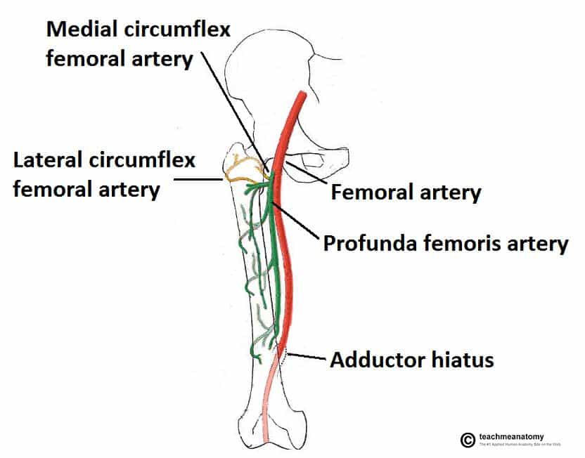
The profunda femoris artery arises from the posterolateral aspect of the femoral artery proper, approximately 3cm distal to the inguinal ligament. It passes inferiorly and posteriorly into the thigh, along the medial aspect of the femur.
Shortly after its origin, it gives rise to the medial and lateral circumflex femoral arteries.
It travels between the pectineus and adductor longus muscles of the medial thigh compartment, and then between the adductor longus and brevis as it moves inferiorly.
The profunda femoris then pierces the adductor magnus muscle, terminating as a perforating vessel within the thigh.
Pro Feature - Dissection Images


Supply
The profunda femoris artery gives rise to two major vessels and (usually) three perforating vessels, which supply the muscles and skin of the lateral, medial and posterior thigh:
- Lateral circumflex femoral artery – supplies the muscles of the anterolateral thigh (vastus lateralis, tensor fascia lata, lateral aspect of rectus femoris and vastus intermedius) and overlying subcutaneous tissue and skin.
- Medial circumflex femoral artery– supplies the head and neck of the femur, muscles of the medial thigh, and overlying subcutaneous tissue and skin.
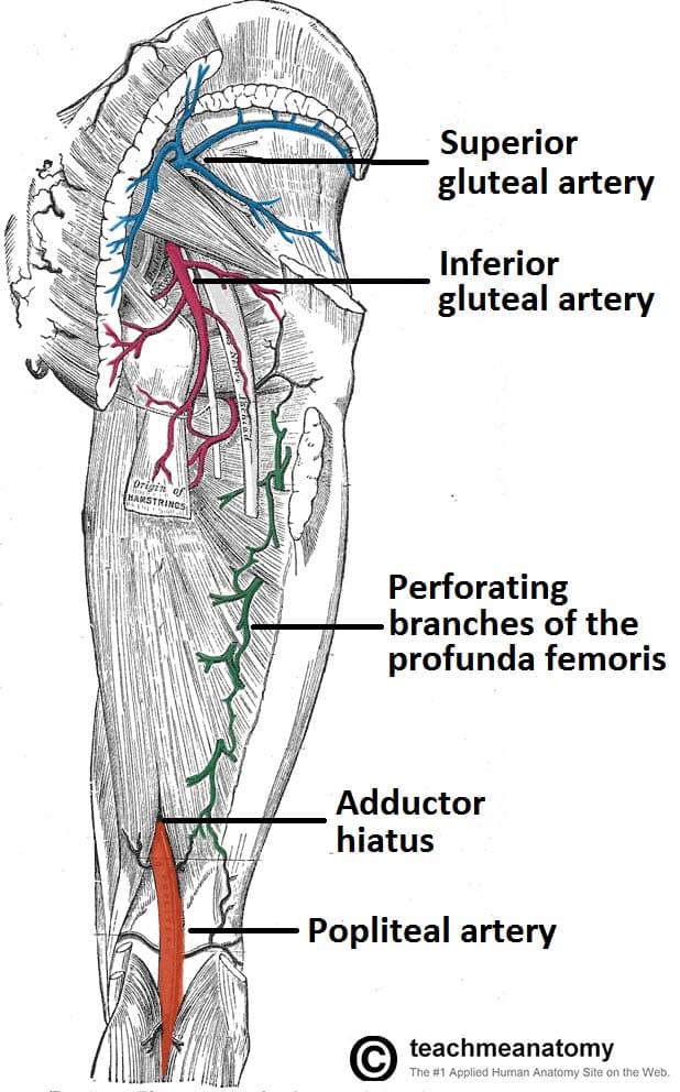
- Perforating arteries (x3) – supplies the muscles of the posterior thigh and overlying subcutaneous tissue and skin.

Fig 2
The arterial supply to the posterior thigh is through perforating branches of the profunda femoris.
