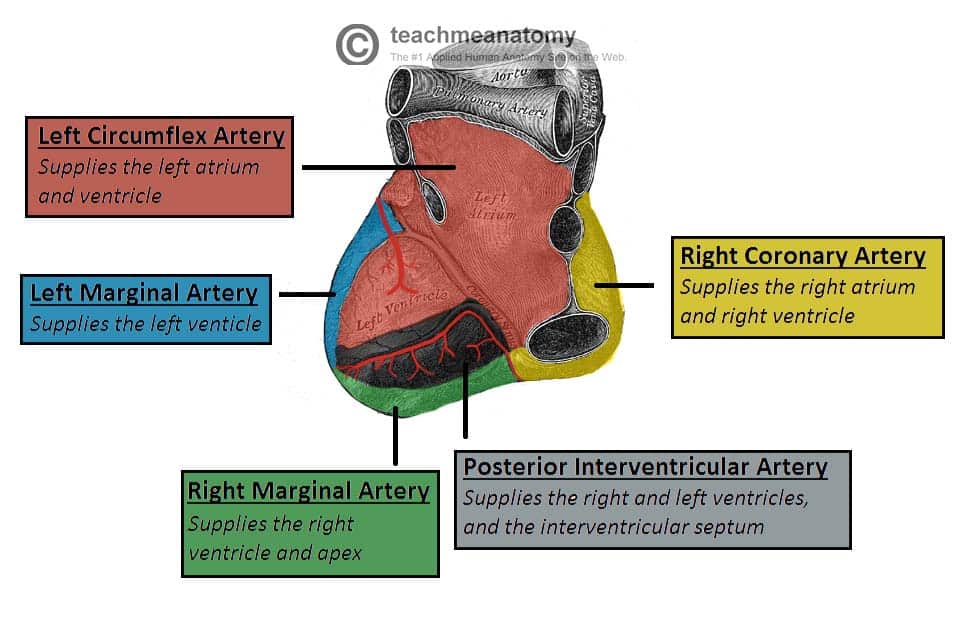
The entire body must be supplied with nutrients and oxygen via the circulatory system and the heart is no exception. The coronary circulation refers to the vessels that supply and drain the heart. Coronary arteries are named as such due to the way they encircle the heart, much like a crown.
This article will outline the naming, distribution, and clinical relevance of vessels in the coronary circulation.
Naming
Coronary Arteries
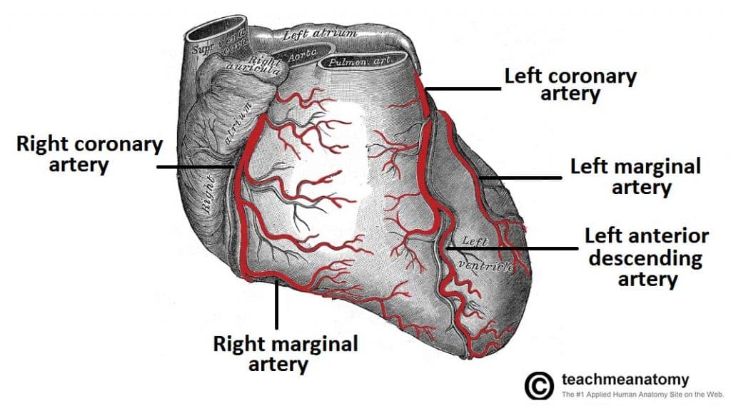
There are two main coronary arteries which branch to supply the entire heart. They are named the left and right coronary arteries, and arise from the left and right aortic sinuses within the aorta.
The aortic sinuses are small openings found within the aorta behind the left and right flaps of the aortic valve. When the heart is relaxed, the back-flow of blood fills these valve pockets, therefore allowing blood to enter the coronary arteries.
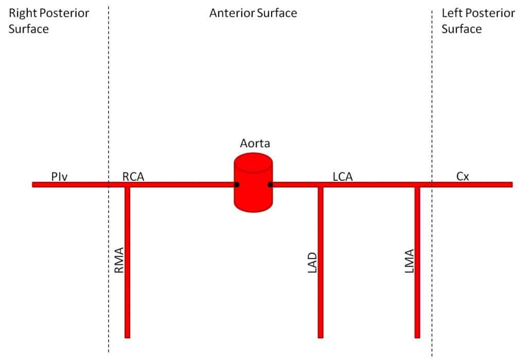
The left coronary artery (LCA) initially branches to yield the left anterior descending (LAD), also called the anterior interventricular artery. The LCA also gives off the left marginal artery (LMA) and the left circumflex artery (Cx). In ~20-25% of individuals, the left circumflex artery contributes to the posterior interventricular artery (PIv).
The right coronary artery (RCA) branches to form the right marginal artery (RMA) anteriorly. In 80-85% of individuals, it also branches into the posterior interventricular artery (PIv) posteriorly.
Cardiac Veins
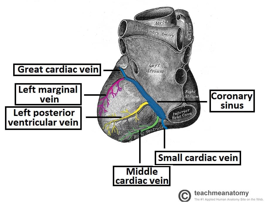
The venous drainage of the heart is mostly through the coronary sinus – a large venous structure located on the posterior aspect of the heart. The cardiac veins drain into the coronary sinus, which in turn, empties into the right atrium. There are also smaller cardiac veins which pass directly into the right atrium.
The main tributaries of the coronary sinus are:
- Great cardiac vein (anterior interventricular vein) – the largest tributary of the coronary sinus. It originates at the apex of the heart and ascends in the anterior interventricular groove. It then curves to the left and continues onto the posterior surface of the heart. Here, it gradually enlarges to form the coronary sinus.
- Small cardiac vein – located on the anterior surface of the heart, in a groove between the right atrium and right ventricle. It travels within this groove onto the posterior surface of the heart, where it empties into the coronary sinus.
- Middle cardiac vein (posterior interventricular vein) – begins at the apex of the heart and ascends in the posterior interventricular groove to empty into the coronary sinus.
- Posterior cardiac vein – located on the posterior surface of the left ventricle. It lies to the left of the middle cardiac vein and empties into the coronary sinus.
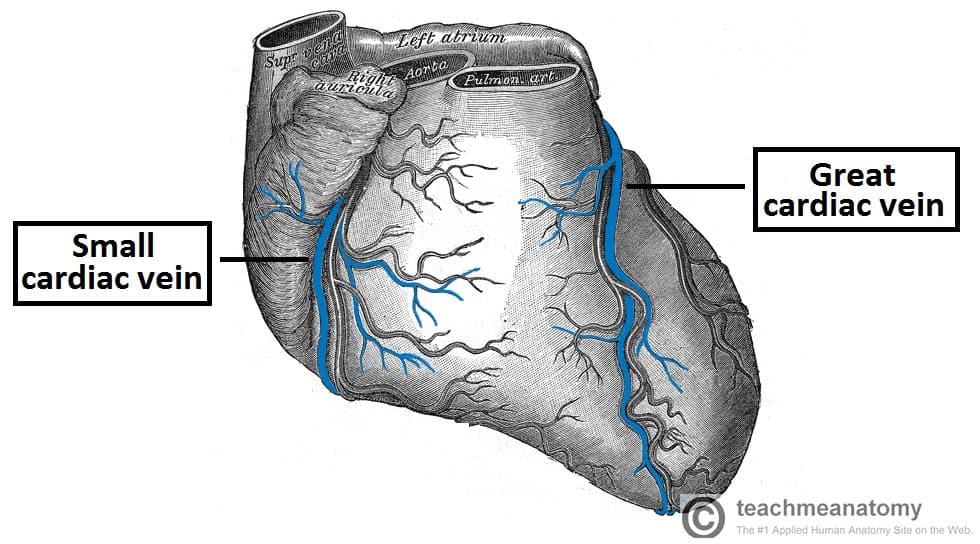
Fig 3 – Anterior view of the venous drainage of the heart. Supplied by the great and small cardiac veins
Distribution of the Coronary Arteries
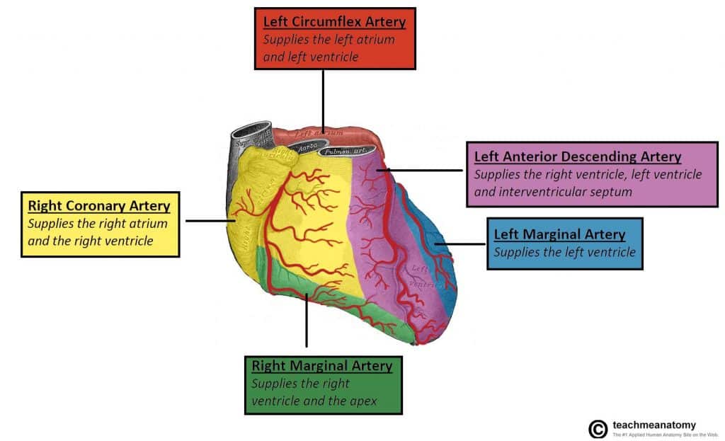
In general, the area of the heart which an artery passes over will be the area that it perfuses. The following describes the anatomical course of the coronary arteries. See Appendix A for a tabular overview of the arterial distribution.
The RCA passes to the right of the pulmonary trunk and runs along the coronary sulcus before branching. The right marginal artery arises from the RCA and moves along the right and inferior border of the heart towards the apex. The RCA continues to the posterior surface of the heart, still running along the coronary sulcus. The posterior interventricular artery then arises from the RCA and follows the posterior interventricular groove towards the apex of the heart.
The LCA passes between the left side of the pulmonary trunk and the left auricle. The LCA divides into the anterior interventricular branch and the circumflex branch. The anterior interventricular branch (LAD) follows the anterior interventricular groove towards the apex of the heart where it continues on the posterior surface to anastomose with the posterior interventricular branch. The circumflex branch follows the coronary sulcus to the left border and onto the posterior surface of the heart. This gives rise to the left marginal branch which follows the left border of the heart.
Clinical Relevance: Coronary Artery Disease
Coronary artery disease or coronary heart disease (CHD) is a leading cause of death, both in the UK and worldwide. It describes a reduction in blood flow to the myocardium and has several causes and consequences.
CHD can result in reduced blood flow to the heart as a result of narrowing or blockage of the coronary arteries. This may be due to atherosclerosis, thrombosis, high blood pressure, diabetes or smoking. All these factors lead to a reduced flow of blood to the heart through physical obstruction or changes in the vessel wall.
Angina pectoris is one consequence of CHD. Angina pectoris describes the transient pain a person may feel on exercise as a result of lack of oxygen supplied to the heart. This pain is felt across the chest but is quickly resolved upon rest. Exercise is a trigger for angina as the coronary arteries fill during the diastolic period of the cardiac cycle. On exercising, the diastolic period is shortened meaning that there is less time for blood flow to overcome a blockage in one of the coronary vessels in order to supply the heart.
If left untreated, angina can soon progress to more severe consequences, such as a myocardial infarction. The sudden occlusion of an artery results in infarction and necrosis of the myocardium. This means a section of the heart is unable to beat (which part of the heart depends on which artery has become occluded). The ECG leads on which an MI change appears can be used to locate the artery that had been occluded as shown in the table.
| Description | ECG leads with changes | Artery occluded |
| Inferior | II, III, aVF | RCA |
| Anteroapical | V3 and V4 | Distal LAD |
| Anteroseptal | V1 and V2 | LAD |
| Anterolateral | I, aVL, V5 and V6 | Circumflex artery |
| Extensive anterior | I, aVL, V2-V6 | Proximal LCA |
| True posterior | Tall R in V1 | RCA |
Diagnosis and Treatment of Coronary Artery Disease
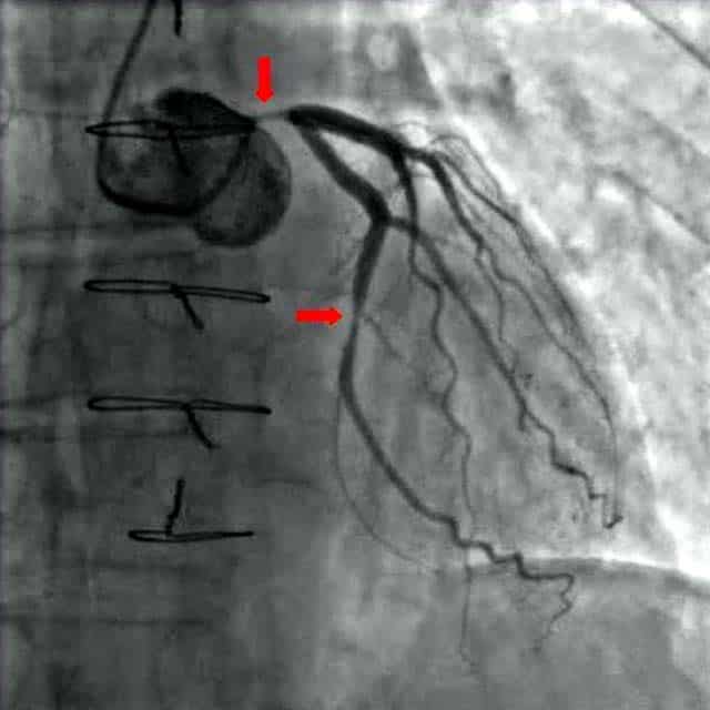
Fig 1.6 – A coronary angiogram. Two critical narrowings have been labelled.
A blockage in a coronary artery can be rapidly identified by performing a coronary angiogram. The imaging modality involves the insertion of a catheter into the aorta via the femoral artery. A contrast dye is injected into the coronary arteries and x-ray based imaging is then used to visualise the coronary arteries and any blockage that may be present.
Immediate treatment of a blockage can be performed by way of a coronary angioplasty, which involves the inflation of a balloon within the affected artery. The balloon pushes aside the atherosclerotic plaque and restores the blood flow to the myocardium. The artery may then be supported by the addition of an intravascular stent to maintain its volume.
Appendix A – Tabular Overview of the Vasculature of the Heart
| Artery | Region supplied | Vein draining region |
| Right coronary | Right atrium
SA and AV nodes Posterior part of interventricular septum (IVS) |
Small cardiac vein
Middle cardiac vein |
| Right marginal | Right ventricle
Apex |
Small cardiac vein
Middle cardiac vein |
| Posterior interventricular | Right ventricle
Left ventricle Posterior 1/3 of IVS |
Left posterior ventricular vein |
| Left coronary | Left atrium
Left ventricle IVS AV bundles |
Great cardiac vein |
| Left anterior descending | Right ventricle
Left ventricle Anterior 2/3 IVS |
Great cardiac vein |
| Left marginal | Left ventricle | Left marginal vein
Great cardiac vein |
| Circumflex | Left atrium
Left ventricle |
Great cardiac vein |
