The muscles of the anterior compartment of the thigh are a group of muscles that (mostly) act to extend the lower limb at the knee joint.
They are collectively innervated by the femoral nerve (L2-L4), and recieve arterial supply from the femoral artery.
In this article, we shall look at the anatomy of the muscles of the anterior thigh – their actions, attachments and clinical correlations.
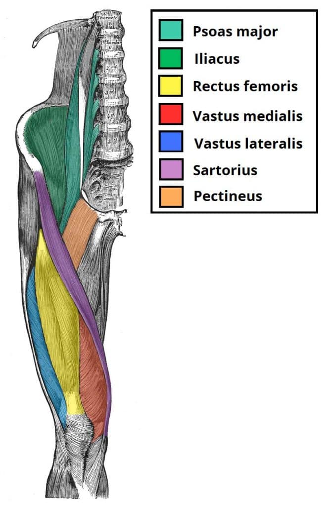
Iliopsoas
The iliopsoas is comprised of two separate muscles; the psoas major and iliacus.
These muscles arise in the pelvis and pass under the inguinal ligament into the anterior compartment of the thigh – where they form a common tendon.
Unlike many of the anterior thigh muscles, the iliopsoas does not perform extension of the leg at the knee joint.
- Attachments: The psoas major originates from the lumbar vertebrae, and the iliacus originates from the iliac fossa of the pelvis. They insert together onto the lesser trochanter of the femur.
- Actions: Flexion of the the thigh at the hip joint.
- Innervation: The psoas major is innervated by anterior rami of L1-3, while the iliacus is innervated by the femoral nerve.
Quadriceps Femoris
The quadriceps femoris consists of four individual muscles – the three vastus muscles and the rectus femoris. It forms the main bulk of the anterior thigh, and is one of the most powerful muscles in the body.
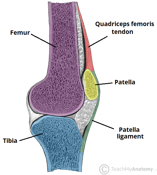
The four muscles collectively insert onto the patella via the quadriceps tendon. The patella, in turn, is attached to the tibial tuberosity by the patella ligament.
Vastus Lateralis
- Proximal attachment: Originates from the greater trochanter and the lateral lip of linea aspera of the femur.
- Actions: Extension of the knee joint. It has a secondary function of stabilising the patella.
- Innervation: Femoral nerve.
Vastus Intermedius
- Proximal attachment: Originates from the anterior and lateral surfaces of the femoral shaft.
- Actions: Extension of the knee joint. It has a secondary function of stabilising the patella.
- Innervation: Femoral nerve.
Vastus Medialis
- Proximal attachment: Originates from the intertrochanteric line and medial lip of the linea aspera of the femur.
- Actions: Extension of the knee joint. It has a secondary function of stabilising the patella.
- Innervation: Femoral nerve.
Rectus Femoris
- Attachments: Originates from the anterior inferior iliac spine and the ilium of the pelvis. It attaches to the patella via the quadriceps femoris tendon.
- Actions: Extension of the knee joint and flexion of the hip joint (it is the only muscle of the quadriceps group to cross both the hip and knee joints).
- Innervation: Femoral nerve.
Sartorius
The sartorius is the longest muscle in the body. It is long and thin, running across the thigh in a inferomedial direction. The sartorius is positioned more superficially than the other muscles in the leg.
- Attachments: Originates from the anterior superior iliac spine, and attaches to the superior, medial surface of the tibia.
- Actions: At the hip joint, it is a flexor, abductor and lateral rotator. At the knee joint, it is also a flexor.
- Innervation: Femoral nerve.

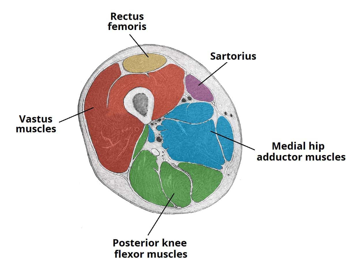
Fig 3 – Cross section of the distal thigh. The iliopsoas and pectineus muscles originate and attach in the proximal thigh, and hence are not included in this diagram.
Pectineus
The pectineus is a flat, quadrangular-shaped muscle which contributes to the floor of the femoral triangle.
- Attachments: Originates from the pectineal line of the pubis bone. It inserts onto the pectineal line on the posterior aspect of the femur, immediately inferior to the lesser trochanter.
- Actions: Adduction and flexion at the hip joint.
- Innervation: Femoral nerve. May also receive a branch from the obturator nerve.
Clinical Relevance: Testing the Quadriceps Femoris
The quadriceps femoris muscle can be used to test the femoral nerve in cases of suspected nerve palsy.
This is performed by positioning the patient supine, with the knee slightly flexed. The patient is asked to extend the leg (at the knee) against resistance. If the femoral nerve is damaged, contraction of the quadriceps femoris will be absent.

