The sublingual glands are the smallest of the three paired salivary glands and the most deeply situated.
Both glands contribute to only 3-5% of overall salivary volume, producing mixed secretions which are predominately mucous in nature. These secretions are important in lubricating food, keeping the oral mucosa moist and initial digestion.
In this article, we shall look at the anatomy of the sublingual glands – their location, blood supply and innervation.
Premium Feature
3D Model
Anatomical Position
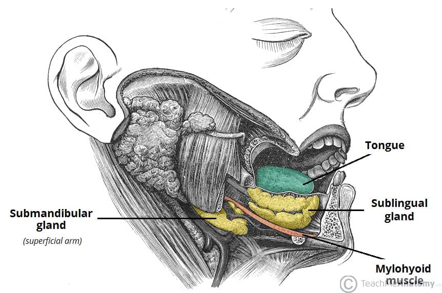
The sublingual glands are almond-shaped and lie on the floor of the oral cavity. They are situated underneath the tongue, bordered laterally by the mandible and medially by genioglossus muscle of the tongue. The glands form a shallow groove on the medial surface of the mandible known as the sublingual fossa.
The submandibular duct and lingual nerve pass alongside the medial aspect of the sublingual gland.
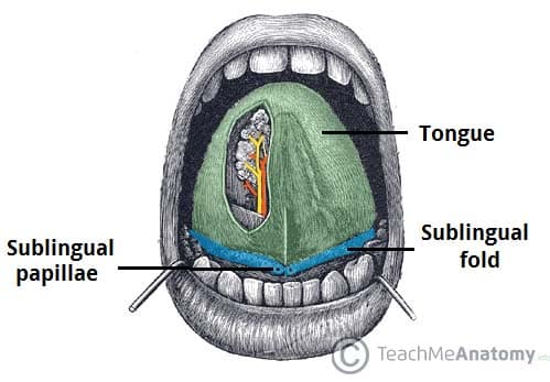
Both sublingual glands unite anteriorly and form a single mass through a horseshoe configuration around the lingual frenulum. The superior aspect of this U-shape forms an elevated, elongate crest of mucous membrane called the sublingual fold (plica sublingualis). Each sublingual fold extends from a posterolateral position and traverses anteriorly to join the sublingual papillae at the midline, either side of the lingual frenulum.
Secretions drain into the oral cavity by minor sublingual ducts (of Rivinus), of which there are 8-20 excretory ducts per gland, each opening out onto the sublingual folds. Through anatomical variance, a major sublingual duct (of Bartholin) can be present in some people. This large accessory duct arises from the inferior aspect of the sublingual gland and then adheres to the passing submandibular duct on its medial side. Drainage then follows the submandibular duct out through the sublingual papillae.
Vasculature
Blood supply is via the sublingual and submental arteries which arise from the lingual and facial arteries respectively; both of the external carotid artery.
Venous drainage is through the sublingual and submental veins which drain into the lingual and facial veins respectively; both then draining into the internal jugular vein.
Innervation
The sublingual glands receive autonomic innervation through parasympathetic and sympathetic fibres, which directly and indirectly regulate salivary secretions respectively. Their innervation is the same as that of the submandibular glands.
Parasympathetic
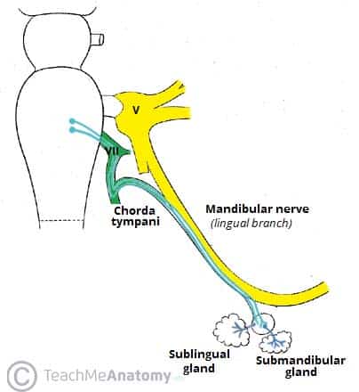
Parasympathetic innervation originates from the superior salivatory nucleus through pre-synaptic fibres via the chorda tympani branch of the facial nerve (CNVII). The chorda tympani then unifies with the lingual branch of the mandibular nerve (CNViii) before synapsing at the submandibular ganglion and suspending it by two nerve filaments.
Post-ganglionic innervation consists of secretomotor fibres which directly induce the gland to produce secretions, and vasodilator fibres which accompany arteries to increase blood supply to the gland. Increased parasympathetic drive promotes saliva secretion.

Fig 3
Innervation to the sublingual gland, via the submandibular ganglion.
Sympathetic
Sympathetic innervation originates from the superior cervical ganglion, where post-synaptic vasoconstrictive fibres travel as a plexus on the internal and external carotid arteries, facial artery and finally the sublingual and submental arteries to enter each gland.
Increased sympathetic drive reduces glandular bloodflow through vasoconstriction and decreases the volume of salivary secretions, resulting in a more mucus saliva.
Clinical Relevance
Ranula
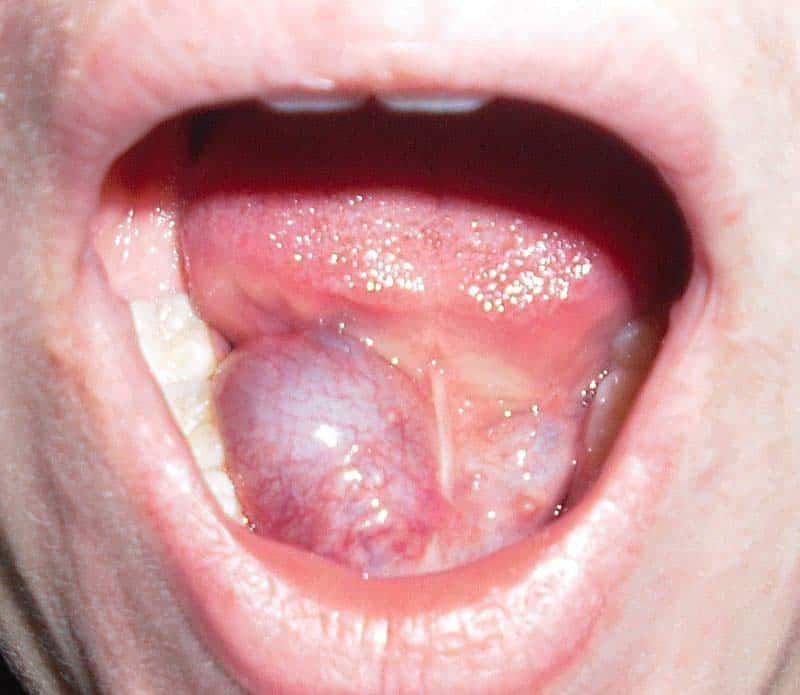
A ranula is a type of mucocele (mucous cyst) that occurs in the floor of the mouth inferior to the tongue. It is the most common disorder associated with the sublingual glands due to their higher mucin content in secretions compared to other salivary glands.
Ranulas can be caused by trauma to the delicate sublingual gland ducts causing them to rupture, with mucin then collecting within the connective tissues to form a cyst.
Ranulas may be small and asymptomatic and can therefore be left alone. Alternatively, they may cause pain and grow large enough to fill the mouth causing dysphagia; an indication for sublingual gland excision. Spilt mucin from the gland or ducts may collect inferiorly beneath mylohyoid and present as a swelling in the neck (a cervical ranula). Rarely, this collection can course posteriorly into the parapharyngeal space.


