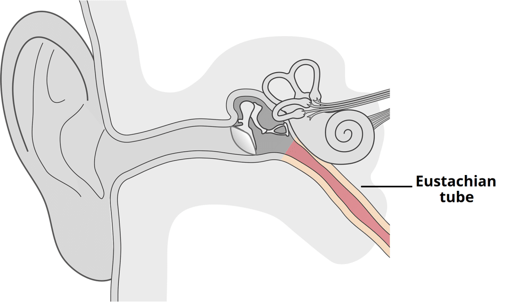
The Eustachian tube (auditory or pharyngotympanic tube) is a canal that connects the tympanic cavity (of the middle ear) to the nasopharynx. It is derived from the embryonic first pharyngeal pouch.
It is a distinct organ which plays several roles in auditory physiology.
- Aeration of the middle ear – creating equal air pressure on both sides of the tympanic membrane.
- Clearance of secretions – permitting movement of mucus from middle ear into the nasopharynx.
- Protection of the middle ear – inhibiting pathogen-laden secretions and sounds created in the nasopharynx from entering the middle ear.
In this article, we shall look at the anatomy of the Eustachian tube – its components, muscles, innervation and clinical correlations.
Parts of the Eustachian Tube
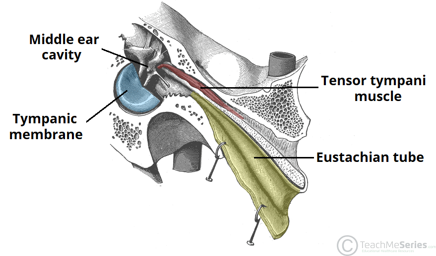
The Eustachian tube arises from the anterior wall of the middle ear and slopes downward, forward, and medially to reach the nasopharynx.
It contains a tympanic and a pharyngeal orifice and is divided into an osseous and cartilaginous portions.
Tympanic Orifice
The tympanic orifice of the Eustachian tube marks the opening of the tube into the middle ear.
It is located just above the middle ear floor. Anterolateral to this opening is Huguier’s canal – where the chorda tympani nerve leaves the middle ear.
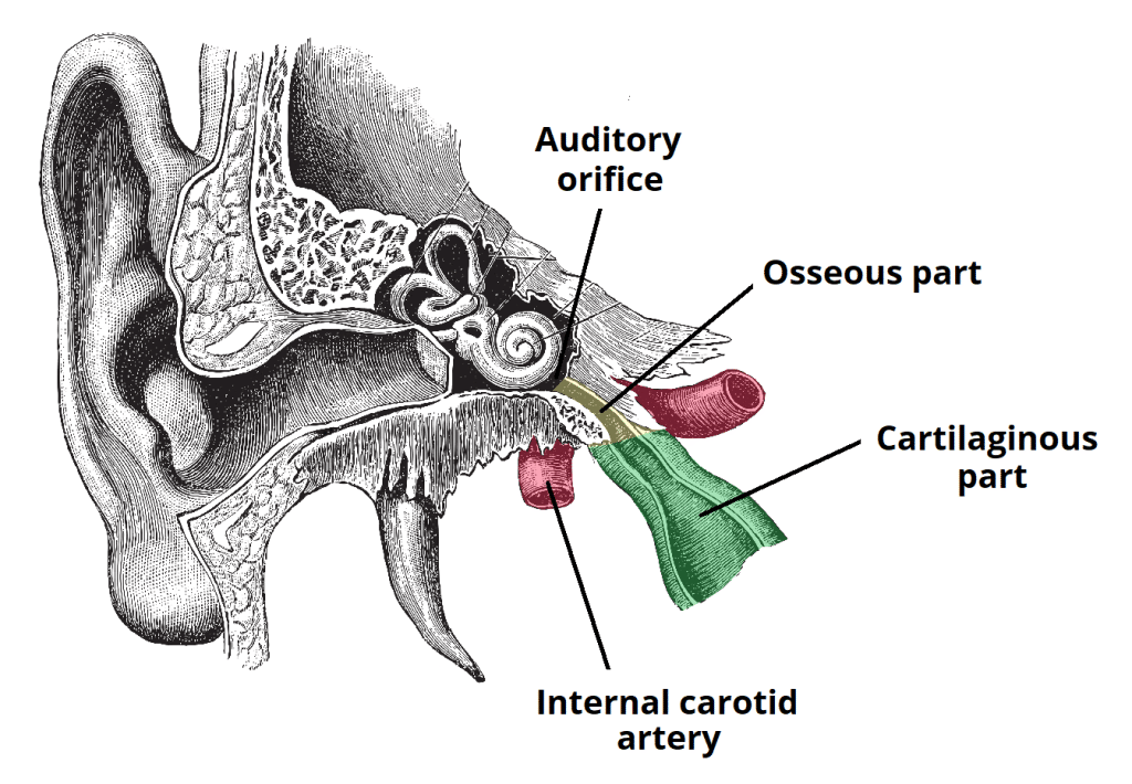
The internal carotid artery is closely associated with the medial wall of the Eustachian tube at the tympanic orifice, where the overlying bone may be thin or even dehiscent in approximately 2% of the cases.
The epithelial lining of the Eustachian tube surrounding the tympanic orifice is ciliated columnar with goblet cells.

Fig 2 – The osseous and cartilaginous parts of the Eustachian tube. Note the proximity of the internal carotid artery to the auditory orifice,
Osseous Portion
The osseous portion refers to the third of the Eustachian tube nearest to the middle ear. It can be variably surrounded by peritubal air cells.
Along the roof of the osseous part of the Eustachian tube is a canal containing the tensor tympani muscle.
The distal end of the osseous portion is formed by the petrous part of the temporal bone. It has a serrated margin for attachment of the cartilaginous component of the tube.
Cartilaginous Portion
The cartilaginous portion refers to the two-thirds of the Eustachian tube nearest to the nasopharynx. It lies in a groove between the petrous part of the temporal bone and the greater wing of the sphenoid bone (petrosphenoidal fissure).
The cartilaginous part is incomplete along the lower and lateral margin of the tube, and is completed by fibrous tissue – the membranous lamina.
Pharyngeal Orifice
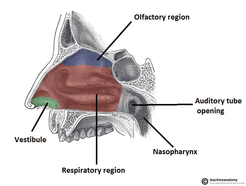
The pharyngeal orifice marks the opening of the Eustachian tube into the nasopharynx. It is located posteriorly to the inferior concha of the nasal cavity.
The epithelial lining of the tube near the pharyngeal orifice is ciliated pseudostratified with goblet cells.

Fig 3 – The opening of the Eustachian tube into the nasopharynx, posterior to the inferior concha.
Associated Muscles
The Eustachian tube is able to passively open when there is a postive pressure difference between the nasopharynx and middle ear (letting air enter or exit).
It also has an active opening mechanism – this is mediated by four muscles surrounding the tube.
| Muscle | Description |
| Tensor Veli Palatini |
|
| Levator Veli Palatini |
|
| Salpingopharyngeus |
|
| Tensor Tympani |
|
Clinical Relevance – Eustachian Tube Dysfunction
The Eustachian tube may be dysfunctional because it is more obstructed or patulous than normal.
An obstructive dysfunction of the tube can be either anatomical or functional. Several conditions can obstruct the tube anatomically. Some are located inside the lumen, like allergy, inflammation, and oedema from gastroesophageal reflux. Others are located outside the lumen, like hypertrophic adenoids and neoplasia of the nasopharynx.
On the other hand, any pathology that affects either the tensor veli palatini muscle itself or its innervation and the geometry of muscle attachment to the cartilaginous part of the tube may result in a functional obstructive dysfunction.
Vasculature
The main arterial supply to the Eustachian tube is from branches of the maxillary artery – the descending palatine and middle meningeal arteries.
There are also contributions from the ascending pharyngeal artery, ascending palatine branch of the facial artery and caroticotympanic branches of the internal carotid artery.
Venous drainage is through the pterygoid venous plexus.
Innervation
The Eustachian tube receives sensory innervation from several nerves:
- Osseous portion – branches of the vagus and glossopharyngeal nerves.
- Cartilaginous portion – branches of the maxillary and mandibular nerves.
Lymphatic Drainage
The lymphatic drainage of the Eustachian tube is into the retropharyngeal, deep jugular, and intraparotid lymph nodes.

