If the heart is the fun, interesting inside bit of an orange, the pericardium could be compared to the peel around it. Like peel, it can seem vaguely unexciting – that is until you learn some of its very important (appeeling. ahem.) physiological functions 1.
In scientific terms, the pericardium is a fibro-serous, fluid-filled sack that surrounds the muscular body of the heart and the roots of the great vessels (the aorta, pulmonary artery, pulmonary veins, and the superior and inferior vena cavae).
This article will give an outline of the functions, structure, innervation, and clinical significance of the pericardium.
Anatomical Structure
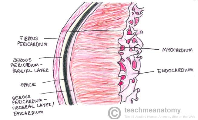
The pericardium is made up of two main layers: a tough external layer known as the fibrous pericardium, and a thin, internal layer known as the serous pericardium (to overextend the orange metaphor, the outer peel could be thought of as the fibrous layer, with the inner white stuff being the serous layer).
Fibrous Pericardium
Continuous with the central tendon of the diaphragm, the fibrous pericardium is made of tough connective tissue and is relatively non-distensible. Its rigid structure prevents rapid overfilling of the heart, but can contribute to serious clinical consequences (see cardiac tamponade).
Serous Pericardium
Enclosed within the fibrous pericardium, the serous pericardium is itself divided into two layers: the outer parietal layer that lines the internal surface of the fibrous pericardium and the internal visceral layer that forms the outer layer of the heart (also known as the epicardium). Each layer is made up of a single sheet of epithelial cells, known as mesothelium.
Found between the outer and inner serous layers is the pericardial cavity, which contains a small amount of lubricating serous fluid. The serous fluid serves to minimize the friction generated by the heart as it contracts.
The order of these layers can be remembered using the acronym Fart Police Smell Villains:
- F – Fibrous layer of the pericardium
- P – Parietal layer of the serous pericardium
- S – Serous fluid
- V – Visceral layer of the serous pericardium
Functions
The pericardium has many physiological roles, the most important of which are detailed below:
- Fixes the heart in the mediastinum and limits its motion. Fixation of the heart is possible because the pericardium is attached to the diaphragm, the sternum, and the tunica adventitia (outer layer) of the great vessels
- Prevents overfilling of the heart. The relatively inextensible fibrous layer of the pericardium prevents the heart from increasing in size too rapidly, thus placing a physical limit on the potential size of the organ
- Lubrication. A thin film of fluid between the two layers of the serous pericardium reduces the friction generated by the heart as it moves within the thoracic cavity
- Protection from infection. The fibrous pericardium serves as a physical barrier between the muscular body of the heart and adjacent organs prone to infection, such as the lungs.

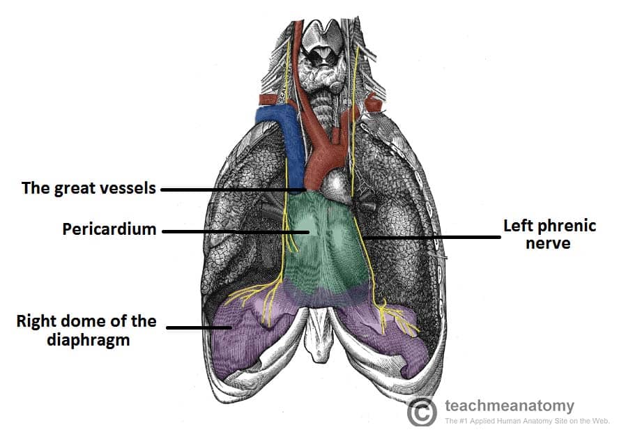
Fig 2 – Anterior view of the pericardium. Note the attachments to the diaphragm, and the roots of the great vessels.
Clinical Relevance: Transverse Pericardial Sinus
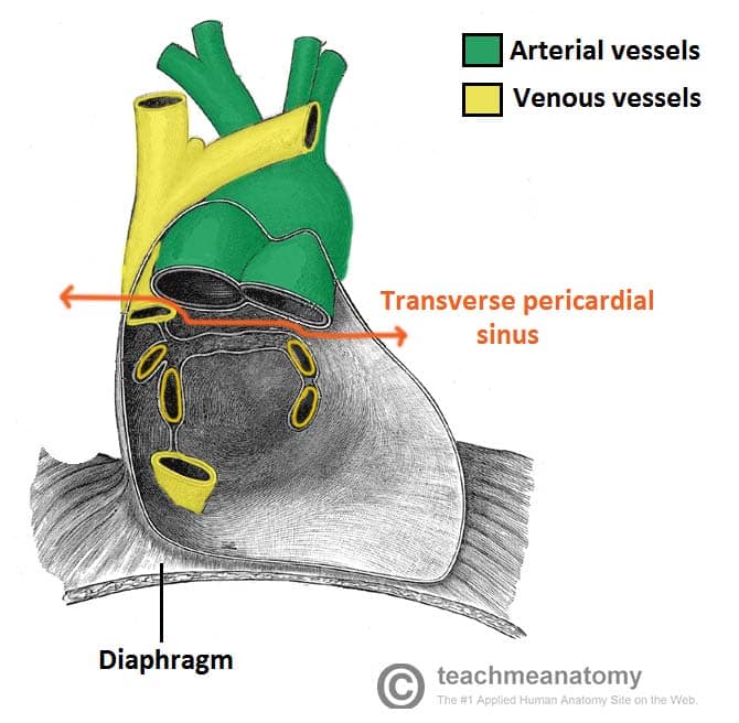
Formed as a result of the embryological folding of the heart tube, the transverse pericardial sinus is a passage through the pericardial cavity.
It is located:
- Posterior to the ascending aorta and pulmonary trunk.
- Anterior to the superior vena cava.
- Superior to the left atrium.
In this position, the transverse pericardial sinus separates the heart’s arterial outflow (aorta, pulmonary trunk) from its venous inflow (superior vena cava, pulmonary veins).
The transverse pericardial sinus can be used to identify and subsequently ligate the arteries of the heart during coronary artery bypass grafting.

Fig 3 – The Transverse pericardial sinus, separating the major arteries and veins. Also note the close relationship of the fibrous pericardium and the diaphragm
Innervation
The phrenic nerve (C3-C5) is responsible for the somatic innervation of the pericardium, as well as providing motor and sensory innervation to the diaphragm. Originating in the neck and travelling down through the thoracic cavity, the phrenic nerve is a common source of referred pain, with a key example being shoulder pain experienced as a result of pericarditis.
Clinical Relevance
Cardiac Tamponade
The relatively inextensible fibrous pericardium can cause problems when there is an accumulation of fluid, known as pericardial effusion, within the pericardial cavity.
The rigid pericardium cannot expand, and thus the heart is subject to the resulting increased pressure. The chambers can become compressed, thus compromising cardiac output.
Pericarditis
Pericarditis, or inflammation of the pericardium, has myriad causes, including bacterial infection and myocardial infarction. The main symptom is chest pain, and the condition can cause acute cardiac tamponade due to an accumulation of fluid in the pericardial cavity.
1 The exciting functions of orange peel are documented at length in academic literature – for instance, did you know that the juice from orange peel is highly flammable?

