The femoral nerve is one of the major peripheral nerves of the lower limb.
In this article, we shall look at the anatomy of the femoral nerve – its anatomical course, functions, and clinical correlations.
Overview
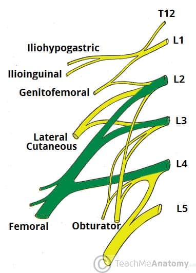
- Nerve roots: L2-L4
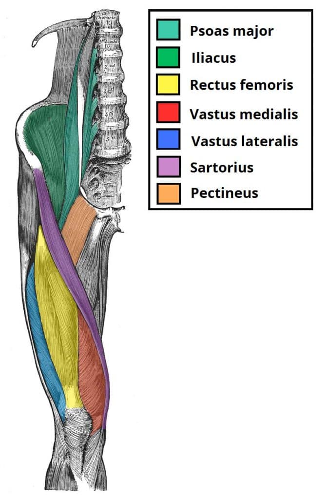
- Motor functions: Innervates the anterior thigh muscles that flex the hip joint (pectineus, iliacus, sartorius) and extend the knee (quadriceps femoris: rectus femoris, vastus lateralis, vastus medialis and vastus intermedius),
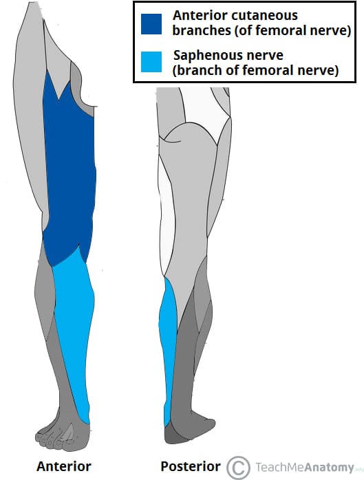
- Sensory functions: Supplies cutaneous branches to the anteromedial thigh (anterior cutaneous branches of the femoral nerve) and the medial side of the leg and foot (saphenous nerve).
Anatomical Course
The femoral nerve is the largest branch of the lumbar plexus. It is derived from the anterior rami of nerve roots L2, L3 and L4.
After arising from the lumbar plexus, the femoral nerve travels inferiorly through the psoas major muscle of the posterior abdominal wall. It supplies branches to the iliacus and pectineus muscles prior to entering the thigh.
The femoral nerve then passes underneath the inguinal ligament to enter the femoral triangle. Within this triangle, the nerve is located lateral to the femoral vessels (unlike the nerve, the femoral artery and vein are enclosed within the femoral sheath).
Approximately 4cm below the inguinal ligament, the femoral nerve divides into anterior and posterior divisions:
| Anterior division of the femoral nerve | Posterior division of the femoral nerve |
|
|
The terminal cutaneous branch of the femoral nerve is the saphenous nerve. It travels through the adductor canal (accompanied by the femoral artery and vein) and exits prior to the adductor hiatus. The saphenous nerve innervates the medial aspect of the leg and the foot.
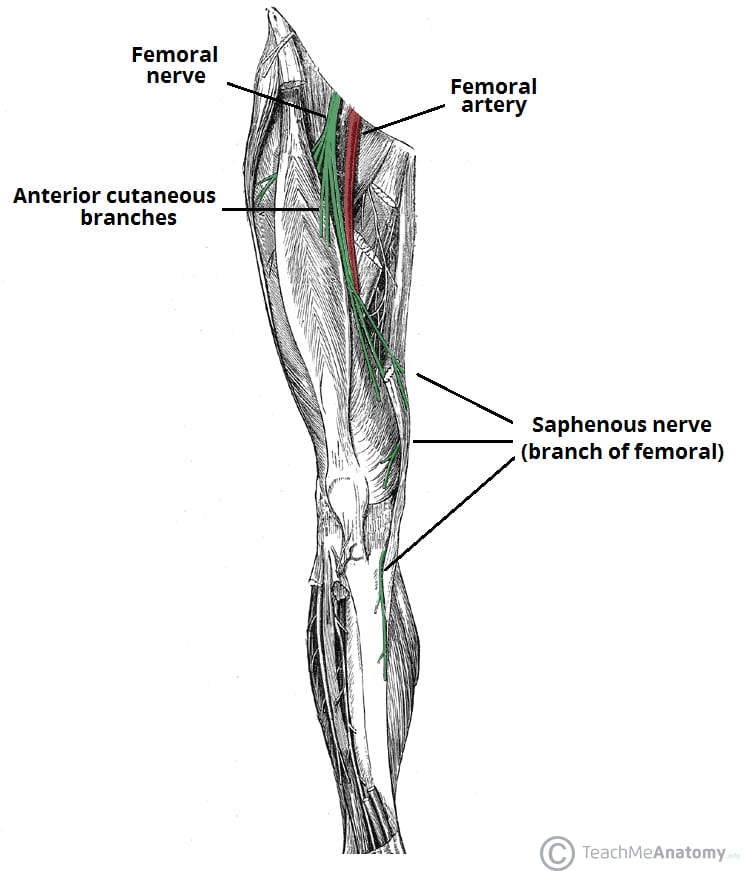
Fig 2 – Anatomical course of the femoral nerve and its two cutaneous branches – anterior cutaneous fibres and saphenous nerve.
Motor Functions
The femoral nerve supplies the muscles of the anterior thigh:
- Hip flexors:
- Pectineus – adducts and flexes the thigh, assists with medial rotation of the thigh.
- Iliacus – acts with psoas major and psoas minor (forming iliopsoas) to flex the thigh at the hip joint and stabilise the hip joint.
- Sartorius – flexes, abducts and laterally rotates the thigh at the hip joint. Flexes the leg at the knee joint.
- Knee extensors:
- Quadriceps femoris (rectus femoris, vastus lateralis, vastus medialis and vastus intermedius) – extends the leg at the knee joint. Rectus femoris also steadies the hip joint and assists iliopsoas in flexing the thigh.
Sensory Functions
There are two main sensory branches that arise from the femoral nerve:
- Anterior cutaneous branches – derived from the anterior division of the femoral nerve. They supply the skin of the anteromedial thigh.
- Saphenous nerve – a continuation of the posterior division of the femoral nerve. It supplies the skin of the medial leg and foot.
Clinical Relevance
Stripping of the Saphenous Vein
The saphenous vein is often stripped in individuals with problematic varicose veins. The long saphenous vein is accompanied in its course by the saphenous nerve.
Damage to the saphenous nerve during this procedure can lead to pain, paraesthesia or complete loss of sensation the medial side of the lower leg.
Femoral Nerve Block
Femoral nerve block (in combination with a sciatic nerve block) may be indicated in patients requiring lower limb surgery who cannot tolerate a general anaesthetic.
A femoral nerve block can also be used as peri- and post-operative analgesia for patients with a fractured neck of femur who cannot tolerate particular analgesics.


