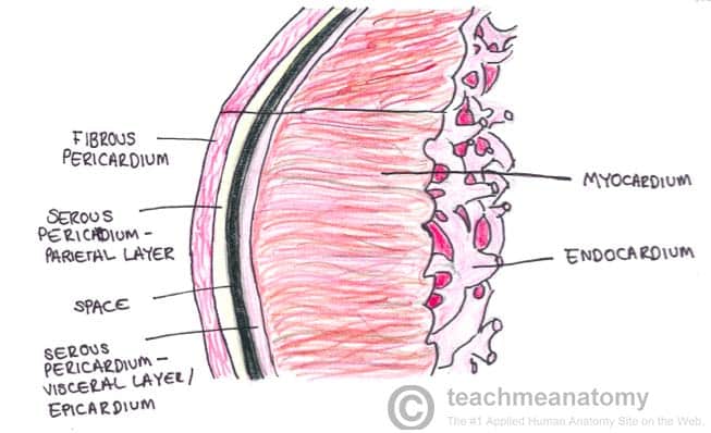
The heart wall itself can be divided into three distinct layers – the endocardium, myocardium, and epicardium.
In this article, we shall look at the anatomy and clinical relevance of these layers.
Endocardium
The innermost layer of the cardiac wall is known as the endocardium. It lines the cavities and valves of the heart.
Structurally, the endocardium is comprised of loose connective tissue and simple squamous epithelial tissue – it is similar in its composition to the endothelium which lines the inside of blood vessels.
In addition to lining the inside of the heart, the endocardium also regulates contractions and aids cardiac embryological development.
Clinical Relevance: Endocarditis
Endocarditis refers to inflammation of the endocardium. It most commonly occurs on the valves of the heart, which the endocardium lines.
The main form of endocarditis is infective endocarditis – caused by a pathogen. Bacteria colonise the heart valve, and cause small clumps of material called vegetations to develop. The resulting inflammation can cause permanent damage to the valve, creating a murmur which is heard when the patient is examined. Furthermore, the damaged valve is more likely to be colonised in the future, resulting in re-infection.
Subendocardial layer
The subendocardial layer lies between, and joins, the endocardium and the myocardium. It consists of a layer of loose fibrous tissue, containing the vessels and nerves of the conducting system of the heart. The purkinje fibres are located in this layer.
As the subendocardial layer houses the conducting system of the heart, damage to this layer can result in various arrhythmias.
Myocardium
The myocardium is composed of cardiac muscle and is an involuntary striated muscle. It is responsible for contractions of the heart.
Clinical Relevance: Myocarditis
Myocarditis refers to an inflammation of the heart muscle, often due to viruses such as adenovirus and coxsackie B. Symptoms depend on the severity of the inflammation, but often include chest pain, shortness of breath, and tachycardia.
The common sequelae of myocarditis is damage to the cardiac muscle of the myocardium. This can result in cardiac arrhythmias and heart failure.
Subepicardial layer
The subepidcardial layer lies between, and joins, the myocardium and the epicardium.
Epicardium
The epicardium is the outermost layer of the heart, formed by the visceral layer of the pericardium. It is composed of connective tissue and fat. The connective tissue secretes a small amount of lubricating fluid into the pericardial cavity.
In addition to the connective tissue and fat, the epicardium is lined by on its outer surface by simple squamous epithelial cells.
