The mastoid fossa (also known as MacEwen’s triangle or suprameatal triangle) is a triangular shaped depression in the external surface of the temporal bone.
It serves as an important anatomical landmark in otologic surgery.
In this article, we shall look at the anatomy of MacEwen’s triangle – its borders, contents and clinical relevance.
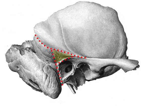
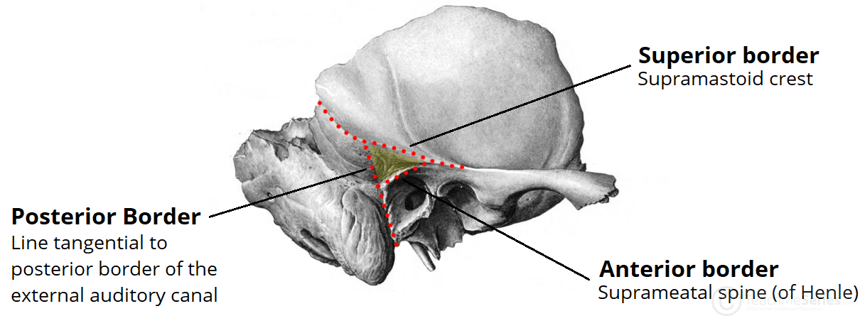
Fig 1 – The mastoid fossa (shaded yellow) is a surface landmark of the external surface of the temporal bone
Borders
The mastoid fossa is a triangular shaped area with superior, anterior, and posterior borders:
- Superior – Supramastoid crest
- Extension of the upper border of the posterior root of the zygomatic process.
- Anterior – Suprameatal spine (spine of Henle)
- Projection of bone at the posterosuperior aspect of the opening of the external acoustic meatus, immediately inferior to the root of the zygomatic process.
- Posterior – Hypothetical vertical line
- Tangential to the mid-point of the posterior wall of the external auditory canal

Fig 2 – The mastoid fossa is triangle shaped. It has superior, anterior, and posterior borders.
Contents
The mastoid fossa is a bony landmark on the external surface of the temporal bone.
It overlies the mastoid antrum – the largest of the mastoid air cells.
The mastoid fossa does not contain any other important structures. This is why it is so significant in otologic surgery – it demarks a ‘safe area‘ to begin the traditional drilling approach during a cortical mastoidectomy.
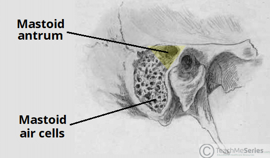
Fig 3 – The mastoid fossa highlighted in yellow with the underlying mastoid antrum.
Clinical Relevance: Cortical Mastoidectomy
A cortical mastoidectomy is a surgical procedure used to remove the mastoid air cells of the temporal bone.
It is typically used to treat severe cases of acute mastoiditis. This is a condition where infection from the middle ear spreads to the mastoid air cells.
The mastoid fossa is an important landmark in a cortical mastoidectomy. It overlies the mastoid antrum, and demarks a ‘safe area‘ to begin the traditional drilling approach.