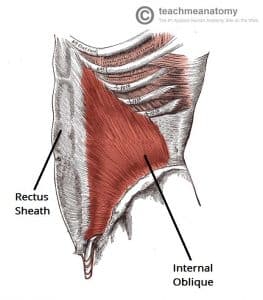
The internal oblique is a muscle of the anterior abdominal wall. It is a broad, sheet-like muscle, located deep to the external oblique.
- Attachments: Originates from the inguinal ligament, iliac crest and lumbodorsal fascia. It inserts onto ribs 10-12.
- Actions: Bilateral contraction compresses the abdomen, while unilateral contraction ipsilaterally rotates the torso.
- Innervation: Thoracoabdominal nerves (T7-T11), subcostal nerve (T12) and branches of the lumbar plexus.
- Blood supply: Lower posterior intercostal and subcostal arteries, superior and inferior epigastric arteries, superficial and deep circumflex arteries, posterior lumbar arteries.
By TeachMeSeries Ltd (2024)

Fig 1 – Lateral view of the abdominal wall. The internal oblique is visible – note that its fibres are perpendicular to those of the external oblique.