The radial nerve is a major peripheral nerve of the upper limb.
In this article, we shall look at the anatomy of the radial nerve – its anatomical course and its motor and sensory functions. We shall also consider the clinical consequences of damage to the nerve.
Overview
- Nerve roots – C5-T1.
- Sensory – Innervates most of the skin of the posterior forearm, the lateral aspect of the dorsum of the hand, and the dorsal surface of the lateral three and a half digits.
- Motor – Innervates the triceps brachii and the extensor muscles in the forearm.
Anatomical Course
The radial nerve is the terminal continuation of the posterior cord of the brachial plexus. It therefore contains fibres from nerve roots C5 – T1.
The nerve arises in the axilla region, where it is situated posteriorly to the axillary artery. It exits the axilla inferiorly (via the triangular interval), and supplies branches to the long and lateral heads of the triceps brachii.
The radial nerve then descends down the arm, travelling in a shallow depression within the surface of the humerus, known as the radial groove.
As it descends, the radial nerve wraps around the humerus laterally, and supplies a branch to the medial head of the triceps brachii. During much of its course within the arm, it is accompanied by the deep branch of the brachial artery.
To enter the forearm, the radial nerve travels anterior to the lateral epicondyle of the humerus, through the cubital fossa. The nerve then terminates by dividing into two branches:
- Deep branch (motor) – innervates the muscles in the posterior compartment of the forearm.
- Superficial branch (sensory) – contributes to the cutaneous innervation of the dorsal hand and fingers.
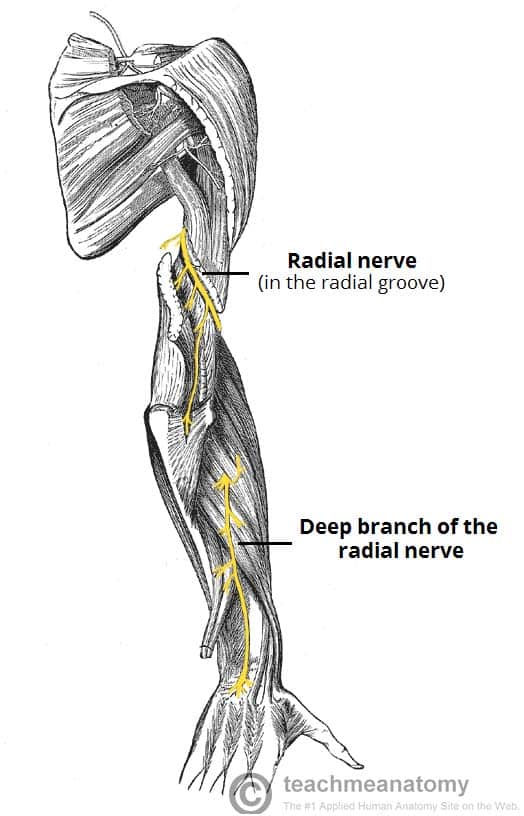
Fig 1 – View of the posterior arm, showing the anatomical course of the radial nerve
Motor Functions
The radial nerve innervates the muscles located in the posterior arm and posterior forearm.
In the arm, it innervates the three heads of the triceps brachii, which acts to extend the arm at the elbow. The radial nerve also gives rise to branches that supply the brachioradialis and extensor carpi radialis longus (muscles of the posterior forearm).
A terminal branch of the radial nerve, the deep branch, innervates the remaining muscles of the posterior forearm. As a generalisation, these muscles act to extend at the wrist and finger joints, and supinate the forearm.
Note: When the deep branch of the radial nerve penetrates the supinator muscle of the forearm, it is termed the posterior interosseous nerve for the remainder of its course.
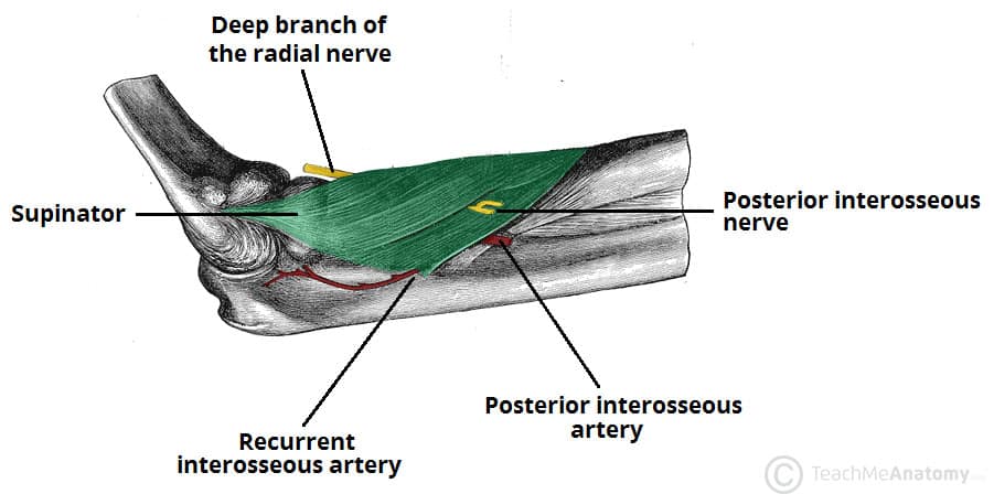
Fig 2 – The deep branch of the radial nerve pierces the supinator muscle, and is renamed the posterior interosseous nerve.
Sensory Functions
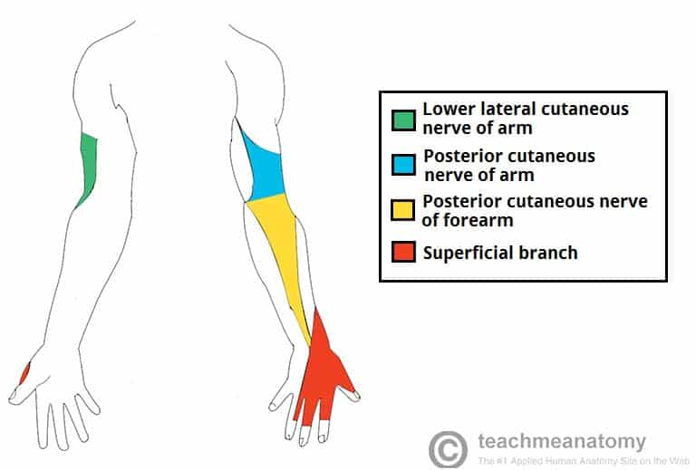
There are four branches of the radial nerve that provide cutaneous innervation to the skin of the upper limb. Three of these branches arise in the upper arm:
- Lower lateral cutaneous nerve of arm – Innervates the lateral aspect of the arm, inferior to the insertion of the deltoid muscle.
- Posterior cutaneous nerve of arm – Innervates the posterior surface of the arm.
- Posterior cutaneous nerve of forearm – Innervates a strip of skin down the middle of the posterior forearm.
The fourth branch – the superficial branch – is a terminal division of the radial nerve. It innervates the dorsal surface of the lateral three and half digits and the associated area on the dorsum of the hand.
Clinical Relevance: Injury to the Radial Nerve
Injury to the radial nerve can be broadly categorised into four groups – depending on where the damage has occurred (and thus which components of the nerve have been affected).
In the Axilla
The radial nerve can be damaged in the axilla region by a dislocation at the shoulder joint, or a fracture of the proximal humerus. Occasionally, it is injured via excessive pressure on the nerve within the axilla (e.g. a badly fitting crutch).
- Motor functions – the triceps brachii and muscles in posterior compartment are affected. The patient is unable to extend at the forearm, wrist and fingers. Unopposed flexion of wrist occurs, known as wrist-drop.
- Sensory functions – all four cutaneous branches of the radial nerve are affected. There will be a loss of sensation over the lateral and posterior arm, posterior forearm, and dorsal surface of the lateral three and a half digits.

Fig 4 – Wristdrop of the left forearm, as a result of radial nerve palsy.
In the Radial Groove
The radial nerve is tightly bound within the spiral groove of the humerus. Thus, it is most susceptible to damage with a fracture of the humeral shaft.
- Motor functions
- The triceps brachii may be weakened, but is not paralysed (branches to the long and lateral heads of the triceps arise proximal to the radial groove).
- Muscles of the posterior forearm are affected. The patient is unable to extend at the wrist and fingers. Unopposed flexion of wrist occurs, known as wrist-drop.
- Sensory functions – the cutaneous branches to the arm and forearm have already arisen. The superficial branch of the radial nerve will be damaged, resulting in sensory loss to the dorsal surface of the lateral three and half digits and the associated area on the dorsum of the hand.
In the Forearm
There are two terminal branches of the radial nerve located within the forearm. The typical mechanism of injury and effect of their injury differs:
| Superficial Branch | Deep Branch | |
| Mechanism | Stabbing or laceration of forearm | Fracture of radial head, or posterior dislocation of radius |
| Motor functions | None | Majority of the muscles in posterior forearm are affected. Wrist-drop does not occur, as the extensor carpi radialis longus is unaffected, and maintains some extension at the wrist |
| Sensory functions | Sensory loss affecting the lateral 3 ½ digits, and associated the associated area on the dorsum of the hand. | None |

