The muscles that act on the hand can be divided into two groups:
- Extrinsic muscles – located in the anterior and posterior compartments of the forearm. They control crude movements and produce a forceful grip.
- Intrinsic muscles – located within the hand itself. They are responsible for the fine motor functions of the hand.
In this article, we shall look at the anatomy of the intrinsic muscles of the hand – their attachments, actions and innervation.
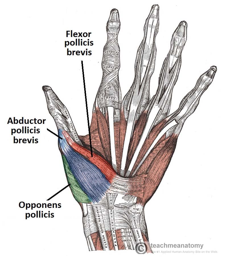
Thenar Muscles
The thenar muscles are three short muscles located at the base of the thumb. The muscle bellies produce a bulge, known as the thenar eminence. They are responsible for the fine movements of the thumb.
The median nerve innervates all the thenar muscles.
Opponens Pollicis
The opponens pollicis is the largest of the thenar muscles, and lies underneath the other two.
- Attachments: Originates from the tubercle of the trapezium and the associated flexor retinaculum. It inserts onto the lateral margin of the first metacarpal.
- Actions: Opposes the thumb, by medially rotating and flexing the metacarpal on the trapezium.
- Innervation: Median nerve (recurrent branch).
Abductor Pollicis Brevis
The abductor pollicis brevis forms the anterolateral aspect of the thenar eminence, overlying the opponens pollicis.
- Attachments: Originates from the tubercles of the scaphoid and trapezium, and from the associated flexor retinaculum. Attaches to lateral side of proximal phalanx of the thumb.
- Actions: Abducts the thumb.
- Innervation: Median nerve (recurrent branch).
Flexor Pollicis Brevis
The flexor pollicis brevis forms the medial aspect of the thenar eminence. It is described as having a superficial and deep part – although the deep component is variable in size.
- Attachments: Originates from the tubercle of the trapezium and from the associated flexor retinaculum. Attaches to the base of the proximal phalanx of the thumb.
- Actions: Flexes the metacarpophalangeal joint of the thumb.
- Innervation: Median nerve (recurrent branch). The deep head is innervated by the deep branch of the ulnar nerve.
Hypothenar Muscles
The hypothenar muscles form the hypothenar eminence – a muscular protrusion on the medial side of the palm, at the base of the little finger. These muscles are similar to the thenar muscles in both name and organisation.
The ulnar nerve innervates the muscles of the hypothenar eminence.
Opponens Digiti Minimi
The opponens digit minimi lies deep to the other hypothenar muscles.
- Attachments: Originates from the hook of hamate and associated flexor retinaculum, inserts into the medial margin of metacarpal V.
- Actions: It rotates the metacarpal of the little finger towards the palm, producing opposition.
- Innervation: Ulnar nerve.
Abductor Digiti Minimi
The abductor digiti minimi is the most superficial of the hypothenar muscle group.
- Attachments: Originates from the pisiform and the tendon of the flexor carpi ulnaris. It attaches to the base of the proximal phalanx of the little finger.
- Actions: Abducts the little finger.
- Innervation: Ulnar nerve.
Flexor Digiti Minimi Brevis
The flexor digiti minimi brevis lies laterally to the abductor digiti minimi in the hand.
- Attachments: Originates from the hook of hamate and adjacent flexor retinaculum, and inserts into the base of the proximal phalanx of the little finger.
- Actions: Flexes the metacarpophalangeal joint of the little finger.
- Innervation: Ulnar nerve.

Lumbricals
There are four lumbricals in the hand, each associated with a finger. They are very crucial to finger movement, linking the extensor tendons to the flexor tendons.
Denervation of these muscles is the basis for the ulnar claw and hand of benediction.
- Attachments: Each lumbrical originates from a tendon of the flexor digitorum profundus. They pass dorsally and laterally around each finger, and inserts into the extensor hood.
- Actions: Flexion at the MCP joint and extension at the interphalangeal (IP) joints of each digit.
- Innervation: The lateral two lumbricals (of the index and middle fingers) are innervated by the median nerve. The medial two lumbricals (of the little and ring fingers) are innervated by the ulnar nerve.
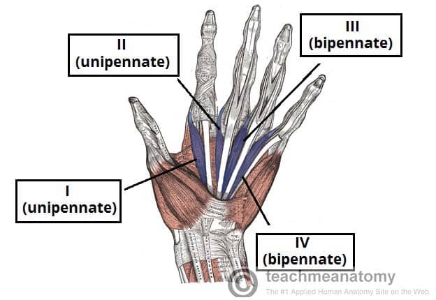
Figure 3 – The lumbricals of the hand. Note the differing unipennate and bipennate structure.
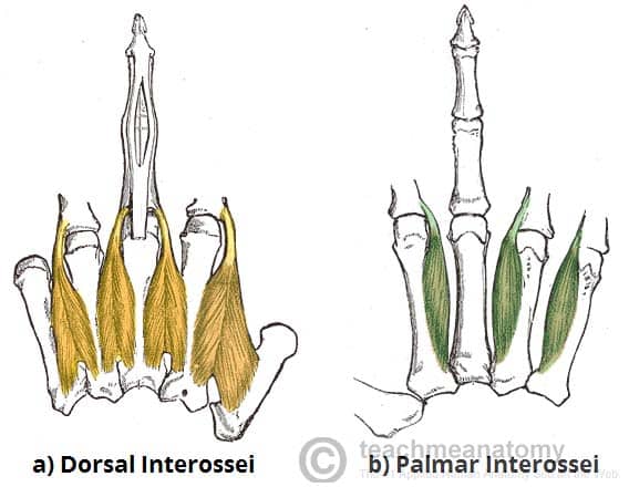
Interossei
The interossei muscles are located between the metacarpal bones of the hand. They can be divided into two groups – dorsal and palmar.
In addition to their actions of abduction (dorsal interossei) and adduction (palmar interossei) of the fingers, the interossei also assist the lumbricals in flexion at the MCP joints and extension at the IP joints.
Dorsal Interossei
The most superficial of all dorsal muscles, these can be palpated on the dorsum of the hand. There are four dorsal interossei muscles.
- Attachments: Each interossei originates from the lateral and medial surfaces of the metacarpals. They attach into the extensor hood and proximal phalanx of each finger.
- Actions: Abduction of the digits. Assists in flexion at the metacarpophalangeal joints and extension at the interphalangeal joints.
- Innervation: Ulnar nerve.
Palmar Interossei
These are located anteriorly on the hand. There are three palmar interossei muscles (although some texts describe a fourth muscle at the base of the proximal phalanx of the thumb).
- Attachments: Each interossei originates from a medial or lateral surface of a metacarpal, and attaches into the extensor hood and proximal phalanx of same finger.
- Actions: Adduction of the digits. Assists in flexion at the metacarpophalangeal joints and extension at the interphalangeal joints.
- Innervation: Ulnar nerve.
Other Muscles in the Palm
There are two other muscles in the palm that are not lumbricals or interossei and do not fit in the hypothenar or thenar compartments:
Palmaris Brevis
The palmaris brevis is a small, thin muscle, found superficially in the subcutaneous tissue of the hypothenar eminence.
- Attachments: Originates from the palmar aponeurosis and flexor retinaculum, attaches to the dermis of the skin on the medial margin of the hand.
- Actions: Wrinkles the skin of the hypothenar eminence and deepens the curvature of the hand, improving grip.
- Innervation: Ulnar nerve.
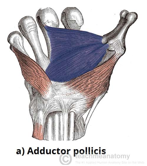
Adductor Pollicis
The adductor pollicis large triangular muscle with two heads. The radial artery passes anteriorly through the space between the two heads, forming the deep palmar arch.
- Attachments: One head originates from metacarpal III. The other head originates from the capitate and adjacent areas of metacarpals II and III. Both attach into the base of the proximal phalanx of the thumb.
- Actions: Adductor of the thumb.
- Innervation: Ulnar nerve.