The trachea, bronchi and bronchioles form the tracheobronchial tree – a system of airways that allow passage of air into the lungs, where gas exchange occurs. These airways are located in the neck and thorax.
In this article we will look at the anatomical position, structure and neurovascular supply of the airways; as well as considering their clinical relevance.
The Trachea
Anatomical Position
The trachea marks the beginning of the tracheobronchial tree. It arises at the lower border of cricoid cartilage in the neck, as a continuation of the larynx.
It travels inferiorly into the superior mediastinum, bifurcating at the level of the sternal angle (forming the right and left main bronchi). As it descends, the trachea is located anteriorly to the oesophagus, and inclines slightly to the right.
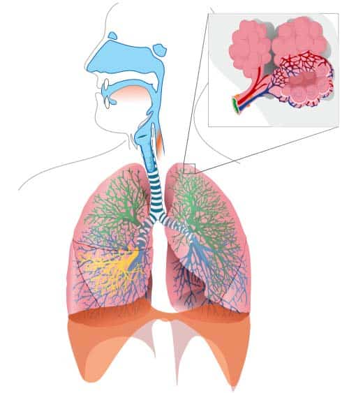
Fig 1 – Overview of the tracheobronchial tree. Key: Green – upper lobe, yellow – middle lobe, blue – lower lobe
Structure
The trachea, like all of the larger respiratory airways, is held open by cartilage – here in C-shaped rings. The free ends of these rings are supported by the trachealis muscle.
The trachea and bronchi are lined by ciliated pseudostratified columnar epithelium, interspersed by goblet cells, which produce mucus. The combination of sweeping movements by the cilia and mucus from the goblet cells forms the functional mucociliary escalator. This acts to trap inhaled particles and pathogens, moving them up out of the airways to be swallowed and destroyed.
At the bifurcation of the primary bronchi, a ridge of cartilage called the carina runs anteroposteriorly between the openings of the two bronchi. This is the most sensitive area of the trachea for triggering the cough reflex, and can be seen on bronchoscopy.
Neurovascular Supply
The trachea receives sensory innervation from the recurrent laryngeal nerve.
Arterial supply comes from the tracheal branches of the inferior thyroid artery, while venous drainage is via the brachiocephalic, azygos and accessory hemiazygos veins.
Bronchi
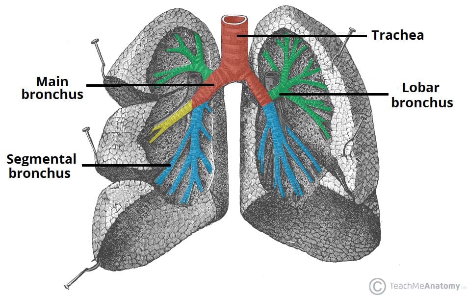
At the level of the sternal angle, the trachea bifurcates into the right and left main bronchi. They undergo further branching to produce the secondary bronchi. Each secondary bronchi supplies a lobe of the lung, and gives rise to several segmental bronchi.
Along with branches of the pulmonary artery and veins, the main bronchi make up the roots of the lungs.
Structure
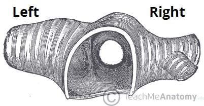
- Right main bronchus – wider, shorter, and descends more vertically than its left-sided counterpart. Clinically, this results in a higher incidence of foreign body inhalation. The right superior lobar bronchus arises before the right main bronchus enters the hilum.
- Left main bronchus – passes inferiorly to the arch of the aorta, and anteriorly to the thoracic aorta and oesophagus in order to reach the hilum of the left lung.
Within the lungs, the main (primary) bronchi branch into lobar (secondary) bronchi. Each secondary bronchi supplies a lobe of the lung, thus there are 3 right lobar bronchi and 2 left. The lobar bronchi then bifurcate into several segmental (tertiary) bronchi, each of which supplies a bronchopulmonary segment. Bronchopulmonary segments are subdivisions of the lung lobes, and act as the functional unit of the lungs.
The structure of bronchi are very similar to that of the trachea, though differences are seen in the shape of their cartilage. In the main bronchi, cartilage rings completely encircle the lumen. However in the smaller lobar and segmental bronchi cartilage is found only in crescent shapes.
Neurovascular Supply
The bronchi derive innervation from pulmonary branches of the vagus nerve (CN X). Blood supply to the bronchi is from branches of the bronchial arteries, while venous drainage is into the bronchial veins.
Bronchioles
The segmental bronchi undergo further branching to form numerous smaller airways – the bronchioles.
Structure
The smallest airways, bronchioles do not contain any cartilage or mucus-secreting goblet cells. Instead, club cells produce a surfactant lipoprotein which is instrumental in preventing the walls of the small airways sticking together during expiration.
Initially there are many generations of conducting bronchioles, which transport air but lack glands and are not involved in gas exchange. Conducting bronchioles then eventually end as terminal bronchioles. These terminal bronchioles branch even further into respiratory bronchioles, which are distinguishable by the presence of alveoli extending from their lumens.
Alveoli are tiny air-filled pockets with thin walls (simple squamous epithelium), and are the sites of gaseous exchange in the lungs. Altogether there are around 300 million alveoli in adult lungs, providing a large surface area for adequate gas exchange.
Clinical Correlations: Asthma
Asthma is a chronic inflammatory disorder of the airways, characterised by hypersensitivity, reversible outflow obstruction and bronchospasm.
There is remodelling of the small airways, causing increased smooth muscle thickness around the bronchioles, damaged epithelium and a thickened basement membrane.
“Asthma attacks” are acute exacerbations of the condition whereby a trigger (e.g. allergens, exercise) causes sudden inflammation and contraction of the smooth muscle around bronchioles (bronchospasm). This narrows the airways, causing difficulty in breathing and wheezing, a characteristic feature of asthma.
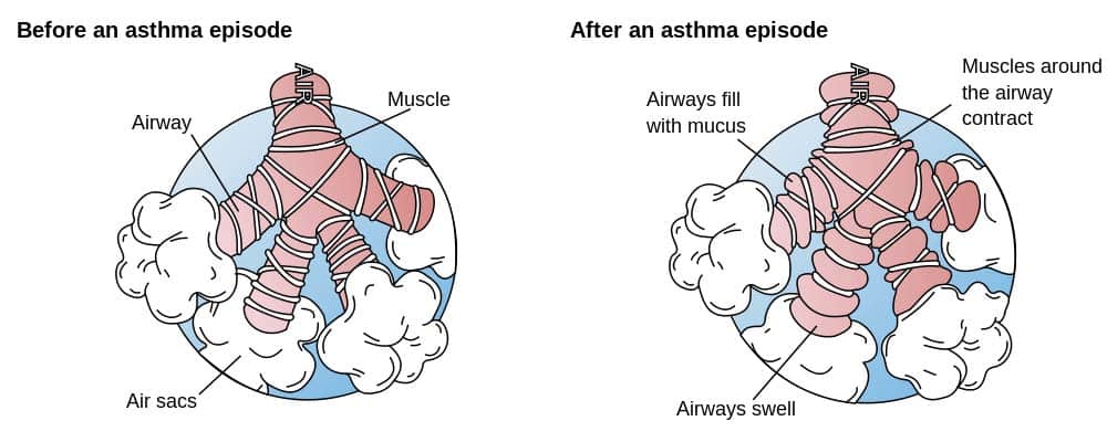
Fig 4 – Diagram showing the effects of an acute asthma exacerbation upon the small airways