The mediastinum is the central compartment of the thoracic cavity, located between the two pleural sacs. It contains most of the thoracic organs, and acts as a conduit for structures traversing the thorax on their way into the abdomen.
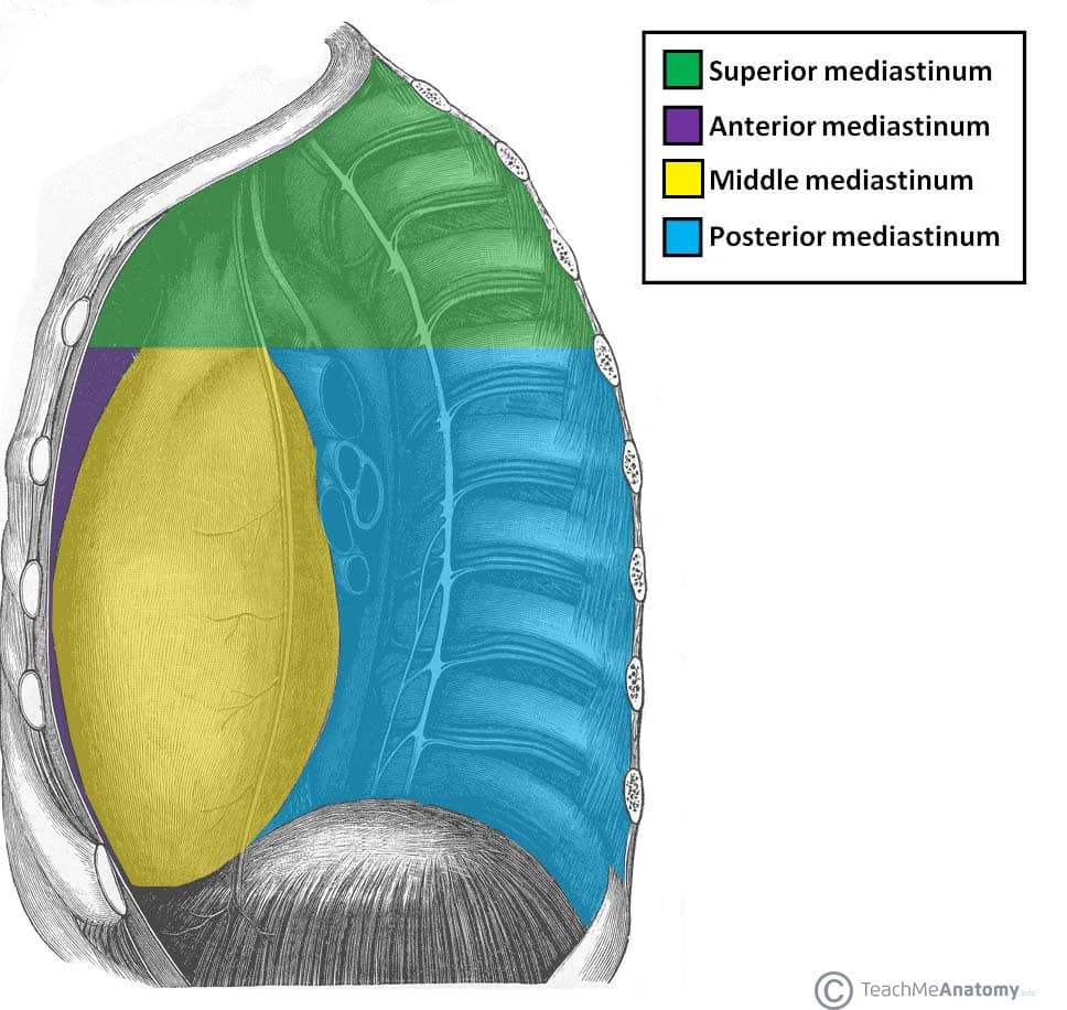
Anatomically, the mediastinum is divided into two parts by an imaginary line that runs from the sternal angle (the angle formed by the junction of the sternal body and manubrium) to the T4 vertebrae:
- Superior mediastinum – extends upwards, terminating at the superior thoracic aperture.
- Inferior mediastinum – extends downwards, terminating at the diaphragm. It is further subdivided into the anterior mediastinum, middle mediastinum and posterior mediastinum.
In this article, we shall look at the anatomy of the posterior mediastinum – its borders, contents and clinical correlations.
Borders
The posterior mediastinum is bordered by the following thoracic structures:
- Lateral: Mediastinal pleura (part of the parietal pleural membrane).
- Anterior: Pericardium.
- Posterior: T5-T12 vertebrae.
- Roof: Imaginary line extending between the sternal angle (the angle formed by the junction of the sternal body and manubrium) and the T4 vertebrae.
- Floor: Diaphragm.
Contents
The posterior mediastinum contains a number of major organs, blood vessels and nerves. We shall now explore the anatomy of these structures in more detail.
Thoracic Aorta
The thoracic (descending) aorta is a continuation of the arch of the aorta, beginning at the lower edge of the T4 vertebra. It descends through the posterior mediastinum to the left of the vertebrae, becoming more medially located as it moves. At the inferior border of T12, the thoracic aorta becomes the abdominal aorta, and passes through the aortic hiatus of the diaphragm.
A number of branches arise from the thoracic aorta in the posterior mediastinum. These tend to arise in three vascular planes; unpaired branches to viscera extend anteriorly, paired branches to viscera extend laterally, and paired segmental parietal branches extend mostly posterolaterally. The major branches are:
- Posterior intercostal arteries – Paired parietal branches. Nine such pairs branch from the posterior aspect of the aorta, supplying the intercostal spaces (except the first two). Pass posteriorly and laterally, in parallel with the ribs.
- Bronchial arteries – Paired visceral branches, usually one or two. The left bronchial arteries always arise directly from the thoracic aorta, while those on the right usually branch indirectly from a right posterior intercostal artery. They go on to supply the tracheobronchial tree.
- Oesophageal arteries – Unpaired visceral branches, arising from the anterior aspect of the aorta. In most individuals there are two, but there can up to five. As the name suggests, these branches go on to supply the oesophagus.
- Superior phrenic arteries – Arise from the anterior aspect of the thoracic aorta at the aortic hiatus, varying in number. They supply the superior aspect of the diaphragm.
Oesophagus
The oesophagus is a muscular tube that connects the pharynx to the stomach; allowing swallowed food to pass into the digestive system. It passes into the posterior mediastinum from the superior mediastinum, descending posteriorly to the arch of the aorta and the heart. Whilst initially positioned to the right, the oesophagus deviates to the left as it moves downwards. It leaves the mediastinum via the oesophageal hiatus of the diaphragm.
The oesophageal plexus is a network of nerves surrounding the oesophagus as it descends, comprising of branches from the left and right vagus nerves. Immediately above the diaphragm, the fibres of the plexus converge to form the anterior vagal trunk and posterior vagal trunk, which travel along the surface of the oesophagus as it exits the thorax.
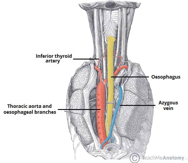
Fig 1.1 – Posterior view of the oesophagus. Some of the thoracic vasculature is noted.
Thoracic Duct
The thoracic duct is the largest lymphatic vessel in the body, allowing return of lymph from most of the body (all but the right superior quadrant) into the venous system.
The duct originates from the cisterna chyli in the abdomen and enters the mediastinum via the aortic hiatus. It ascends to lie directly anterior to the T6-T12 vertebrae, before deviating left as it ascends into the superior mediastinum. While located in the posterior mediastinum, the thoracic duct receives lymphatic drainage from the intercostal spaces and neighbouring anatomical structures through a number of branches.
Azygos System of Veins
This venous network drains blood from the body walls and mediastinal viscera and empties into the superior vena cava. It consists of three major veins:
- Azygos vein – Formed by the union of the right lumbar vein and the right subcostal vein. It enters the mediastinum via the aortic hiatus and drains into the superior vena cava.
- Hemiazygos vein – Formed by the union of the left lumbar vein and left subcostal vein. It enters the mediastinum through the left crus of the diaphragm, ascending on the left side. At the level of T8, it turns to the right and combines with the azygos vein.
- Accessory hemiazygos vein – Formed by the union of the fourth to eighth intercostal veins. It drains into the azygos vein at T7.
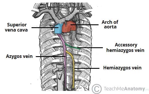
Fig 1.2 – The azygos venous network, which empties into the superior vena cava
Sympathetic Trunks
The sympathetic trunks are paired bundles of nerves that extend from the base of the skull to the coccyx. In the thoracic region, these nerve bundles are known as the thoracic sympathetic trunks. As they descend through the thorax, they lie within the posterior mediastinum.
Arising from these trunks are the lower thoracic splanchnic nerves – they continue inferiorly to supply the viscera of the abdomen.