The ligaments of the female reproductive tract are a series of structures that support the internal female genitalia in the pelvis.
The ligaments of the female reproductive tract can be divided into three categories:
- Broad ligament – a sheet of peritoneum, associated with both the uterus and ovaries.
- Uterine ligaments – ligaments primarily associated with the uterus.
- Ovarian ligaments – ligaments primary associated with the ovaries.
Collectively, these ligaments are tough and non-extensible. They act to support the female viscera and provide a conduit for neurovascular structures.
In this article, we shall look at the attachments and anatomical relations of the ligaments of the female reproductive tract.
Broad Ligament
The broad ligament is a flat sheet of peritoneum, associated with the uterus, fallopian tubes and ovaries. It extends from the lateral pelvic walls on both sides, and folds over the internal female genitalia, covering their surface anteriorly and posteriorly.
Subdivisions
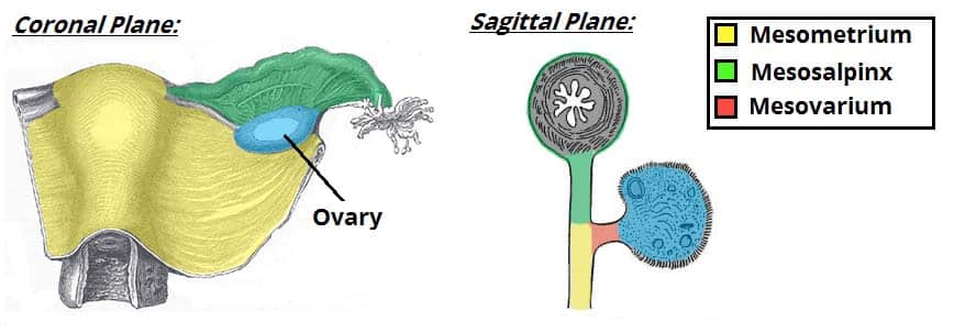
Anatomically, the broad ligament can be divided into three regions:
- Mesometrium – Surrounds the uterus and is the largest subsection of the broad ligament. It runs laterally to cover the external iliac vessels, forming a distinct fold over them. The mesometrium also encloses the proximal part of the round ligament of the uterus.
- Mesovarium – Part of the broad ligament associated with the ovaries. It projects from the posterior surface of the broad ligament and attaches to the hilum of the ovary, enclosing its neurovascular supply. It does not, however, cover the surface of the ovary itself.
- Mesosalpinx – Originates superiorly to the mesovarium, enclosing the fallopian tubes.
Anatomical Relations
The broad ligament is related to many structures within the female pelvis. It is attached to the uterus, fallopian tubes and ovaries. These organs are supplied by the ovarian and uterine arteries, which are also contained within the broad ligament.
Three other ligaments of the female reproductive tract are located within the broad ligament:
- Ovarian ligament.
- Round ligament of uterus.
- Suspensory ligament of ovary (also known as the infundibulopelvic ligament).
(These ligaments shall be explored in more detail later in the article).
Ligaments Associated with the Ovary
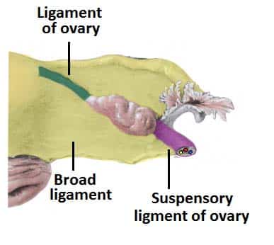
There are two main ligaments that attach to the ovary – the ovarian ligament and suspensory ligament of ovary.
Ovarian Ligament
The ovarian ligament is attached to the ovary inferiorly. It connects the ovary to the side of the uterus. Structurally, it is a fibrous band of tissue that lies within the broad ligament. It joins the uterus just below the origin of the fallopian tubes.
Suspensory Ligament of Ovary
The suspensory ligament of ovary extends outwards from the ovary to the lateral abdominal wall. It consists of a fold of peritoneum, thus some sources consider it to be part of the broad ligament. The function of this ligament is to contain the ovarian vessels and nerves (ovarian artery, ovarian vein, ovarian nerve plexus and lymphatic vessels).
Ligaments Associated with the Uterus
There are a number of ligamentous structures that attach to the uterus. They can be divided by where they attach to the uterus:
- Superior aspect – supported by the broad ligament and the round ligaments.
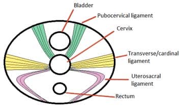
- Middle aspect – supported by the cardinal, pubocervical and uterosacral ligaments.
The inferior aspect of uterus is supported by the structures in the pelvic floor – the levator ani, perineal membrane and perineal body.
Round Ligament
The round ligament is a remnant of the embryonic gubernaculum.
It originates at the uterine horns (the points at which the fallopian tubes enter the uterus), and attaches to the labia majora, passing through the inguinal canal.
The round ligament can be a source of pain during pregnancy, due to the increased force placed on the ligament by the expanding uterus.
Cardinal Ligaments
The cardinal ligaments are also known as the lateral, transverse cervical, or Mackenrodt’s ligaments. They are situated along the inferior border of the broad ligament and house the uterine artery and uterine veins.
These ligaments arise from the side of the cervix and the lateral fornix of the vagina. They provide an extensive attachment on the lateral pelvic wall at the level of the ischial spines. Some fibres of the cardinal ligaments interdigitate with fibres from the uterosacral ligaments.
When a hysterectomy is being performed due to a malignancy, the cardinal ligaments are often removed as they are common reservoir of cancerous cells.
Pubocervical Ligaments
The pubocervical ligaments are bilateral structures, which attach the cervix to the posterior surface of the pubic symphysis. They function to support the uterus within the pelvic cavity.
Uterosacral Ligaments
The uterosacral ligaments are also bilateral fibrous bands, which attach the cervix to the sacrum. They are also known as the recto-uterine ligaments or sacrocervical ligaments. This supports the uterus and holds it in place.
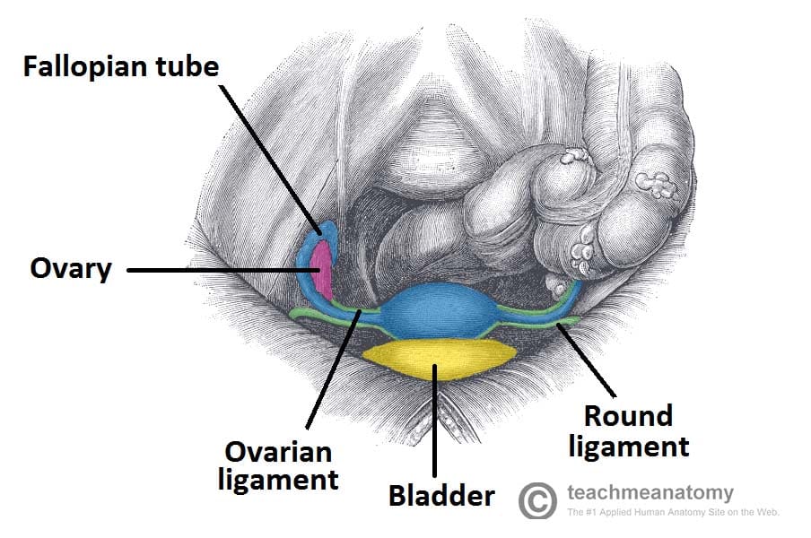
Fig 3 – Overview of the uterus and fallopian tubes, and associated ligaments