The superficial fibular nerve (superficial peroneal nerve) is a nerve of the lower limb.
In this article, we shall look at the anatomy of the superficial fibular nerve – its anatomical course, functions and clinical correlations.
Overview
- Nerve roots: L4-S1
- Motor: Innervates the muscles in the lateral compartment of the leg.
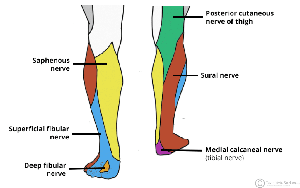
- Sensory: Supplies the anterolateral aspect of the distal leg and the majority of the dorsum of the foot (apart from the webbing between the hallux and the second digit).
Anatomical Course
The superficial fibular nerve is a terminal branch of the common fibular nerve.
It arises at the neck of the fibula, descending between the fibularis muscles and the lateral side of the extensor digitorum longus. Here, it gives rise to motor branches, which supply the fibularis longus and brevis. The nerve continues its descent, with a purely cutaneous function, providing sensory innervation to the anterolateral aspect of the lower leg.
When the superficial fibular nerve reaches the lower third of the leg, it pierces the deep crural fascia and terminates by dividing into the medial and intermedial dorsal cutaneous nerves. These nerves enter the foot to innervate the majority of its dorsal surface.
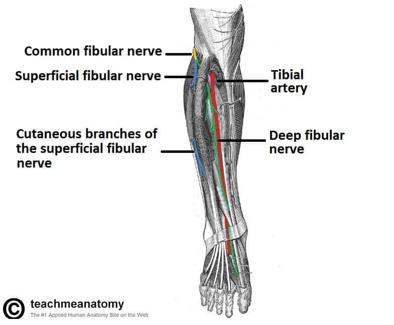
Fig 1 – Anterior view of the leg, showing the major nerves. The proximal portion of the fibularis longus has been removed to show the bifurcation of the common fibular nerve.
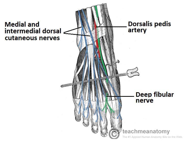
Fig 2 – The cutaneous nerves of the foot. Note the distribution of the dorsal cutaneous nerves
Motor Functions
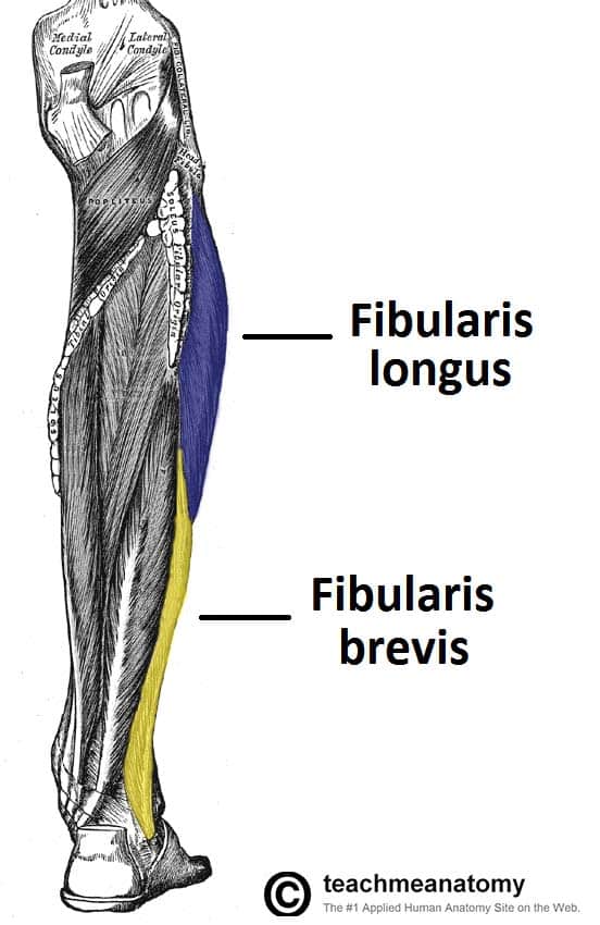
The superficial fibular nerve supplies the fibularis longus and the fibularis brevis. These muscles form the lateral compartment of the leg.
These muscles act to evert the foot (turn outwards) at the subtalar joint. They also weakly contribute to plantarflexion, although this action is mainly carried out by the gastrocnemius and the soleus muscles.
Sensory Functions
The superficial fibular nerve provides cutaneous innervation to certain areas of the leg and foot:
- Innervates the skin over the anterolateral leg, via cutaneous branches directly from the superficial fibular nerve.
- Innervates the skin of the dorsum of the foot (except the first webbed space), via the medial and intermedial dorsal cutaneous nerves.
The dermatomes to which these areas correspond are L5 and S1.
Clinical Relevance
There are two relatively common pathologies involving the damage to the superficial fibular nerve; entrapment and direct damage (e.g from a comminuted fracture).
Superficial Fibular Nerve Entrapment
Superficial peroneal nerve entrapment (also known as nerve compression) can cause pain, and paraesthesia over the lower leg and dorsum of the foot. Entrapment frequently results from ankle sprains or twisting of the ankle, as this causes the nerve to stretch in the lower leg.
Another cause of nerve entrapment occurs at the point where the nerve exits the deep fascia of the leg, the nerve becoming compressed by this fascia. Surgical decompression of the nerve therefore is used to provide relief from the symptoms and pain.
Direct Damage to the Superficial Fibular Nerve
The superficial fibular nerve may be damaged by fracture of the fibula, or by a perforating wound to the lateral side of the leg.
As the muscles that the superficial fibular nerve innervates are evertors, injury to the nerve may result in a loss of eversion. A loss of sensation over the majority of the dorsum of the foot and the anterolateral aspect of the lower leg could also result.
