The obturator nerve is a major peripheral nerve of the lower limb.
In this article, we shall look at the anatomy of the obturator nerve – its anatomical course, functions and clinical correlations.
Overview
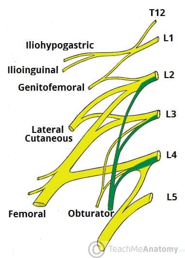
- Nerve roots: L2-L4
- Motor functions: Innervates the muscles of the medial compartment of the thigh (obturator externus, adductor longus, adductor brevis, adductor magnus and gracilis).
- Sensory functions: Cutaneous branches of the obturator nerve innervate the skin of the medial thigh.
Anatomical Course
The obturator nerve is formed from the lumbar plexus. It receives fibres from the anterior divisions of L2, L3 and L4.
After its formation, the obturator nerve descends through the fibres of the psoas major and emerges from its medial border. It then travels posteriorly to the common iliac arteries and laterally along the pelvic wall – towards the obturator foramen of the pelvis.
The obturator nerve enters the medial thigh via the obturator canal (formed within the obturator foramen by the obturator membrane). It then divides into anterior and posterior branches:
- Anterior division (anterior to the adductor brevis):
- Descends in a plane between the adductor longus and adductor brevis (towards the femoral artery).
- Here, it supplies motor fibres to the adductor longus, adductor brevis and gracilis. It can also supply the pectineus muscle.
- It then pierces the fascia lata to become the cutaneous branch of the obturator nerve.
- Posterior division (posterior to the adductor brevis):
- Pierces the obturator externus muscle, and then descends in a plane between the adductor brevis and adductor magnus.
- Innervates the obturator externus and adductor magnus muscles.
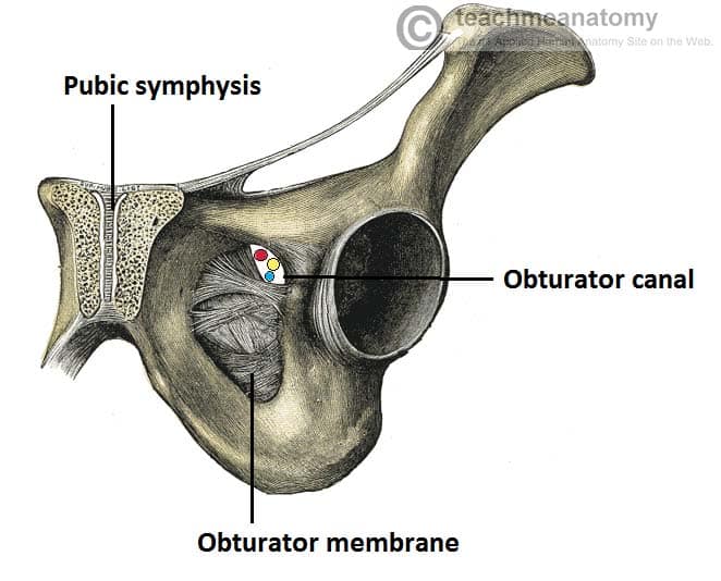
Fig 2 – The obturator canal, formed by the obturator membrane within the obturator foramen of the pelvis.
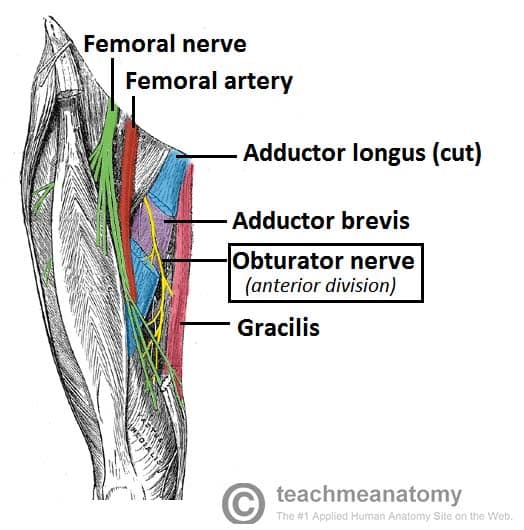
Fig 3 – View of the medial thigh, with the course of the obturator nerve highlighted.
Motor Functions
The obturator nerve innervates all the muscles in the medial compartment of the thigh – except the hamstring part of the adductor magnus (innervated by the tibial nerve).
They are collectively known as the hip adductors:
- Adductor longus – adducts thigh
- Adductor brevis – adducts thigh
- Adductor magnus:
- Adductor part – adducts and flexes thigh
- Hamstring part – extends thigh
- Gracilis – adducts thigh
- Obturator externus – laterally rotates thigh
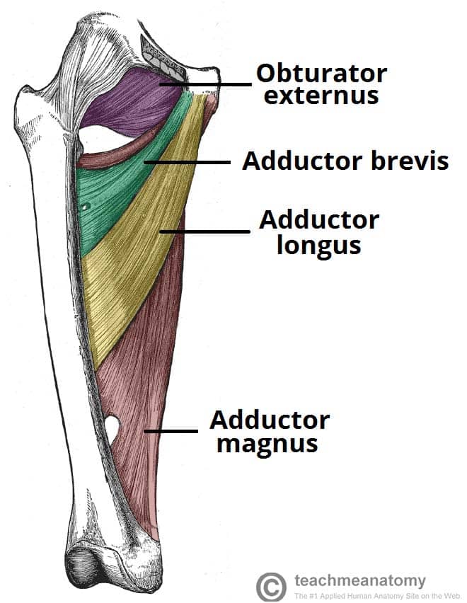
Fig 4 – Muscles of the medial thigh. The overlying muscles in the anterior compartment have been removed.
Sensory Function
The cutaneous branch of the obturator nerve supplies the skin of the middle part of the medial thigh.
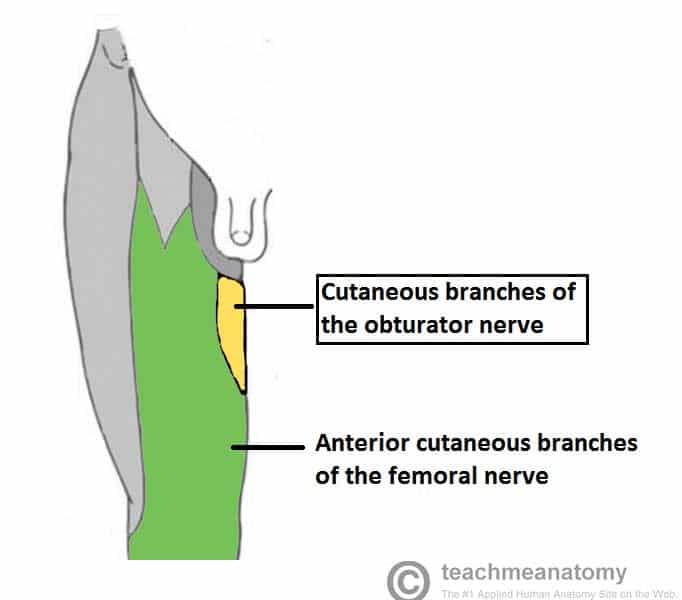
Fig 5 – Image of the cutaneous distribution of the anterior thigh, with the area supplied by the obturator nerve highlighted.
Clinical Relevance
Damage to the Obturator Nerve
The obturator nerve can be damaged during surgery involving the pelvis or abdomen.
Symptoms include numbness and paraesthesia on the medial aspect of the thigh and weakness in adduction of the thigh. Alternatively, the patient could present with posture and gait problems due to the loss of adduction.
Obturator Nerve Block
Obturator nerve block is used in the management of pain after lower limb surgery or for chronic hip pain. The anaesthetic is injected inferior to the pubic tubercle and lateral to the tendon of the adductor longus muscle. This procedure can also be carried out under ultrasound guidance.

