The muscles acting on the foot can be divided into two distinct groups; extrinsic and intrinsic muscles.
- Extrinsic muscles arise from the anterior, posterior and lateral compartments of the leg. They are mainly responsible for actions such as eversion, inversion, plantarflexion and dorsiflexion of the foot.
- Intrinsic muscles are located within the foot and are responsible for the fine motor actions of the foot, for example movement of individual digits.
In this article, we shall examine the anatomy of the intrinsic muscles of the foot. They can be divided into those situated on the dorsum of the foot, and those in the sole of the foot.
Dorsal Aspect
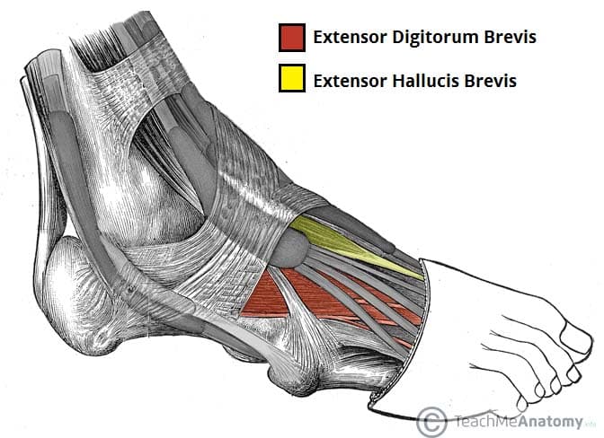
There are two intrinsic muscles located within the dorsum of the foot – the extensor digitorum brevis and extensor hallucis brevis.
They assist the extrinsic muscles of the foot in extending the toes and are both innervated by the deep fibular nerve.
Extensor Digitorum Brevis
The extensor digitorum brevis is a small, thin muscle which lies underneath the long extensor tendons of the foot.
- Attachments: Originates from the calcaneus and inferior extensor retinaculum. It attaches onto the long extensor tendons of toes 2-4.
- Actions: Extension of the lateral four toes.
- Innervation: Deep fibular nerve.
Extensor Hallucis Brevis
The extensor hallucis brevis is often considered to be the medial part of the extensor digitorum brevis muscle, rather than a separate structure.
- Attachments: Originates from the calcaneus and inferior extensor retinaculum. It attaches to the base of the proximal phalanx of the great toe.
- Actions: Extension of the great toe.
- Innervation: Deep fibular nerve.
Plantar Aspect
There are ten intrinsic muscles located in the plantar aspect (sole) of the foot.
They act collectively to stabilise the arches of the foot and individually to control movement of the digits. They are innervated by the medial or lateral plantar nerves – which are branches of the tibial nerve.
The muscles of the plantar aspect are arranged in four layers (superficial to deep):
First Layer
The first layer contains three muscles. It is the most superficial and is located immediately underneath the plantar fascia.
Abductor Hallucis
The abductor hallucis muscle is located on the medial side of the sole, where it contributes to a small soft tissue bulge.
- Attachments: Originates from the medial tubercle of the calcaneus, the flexor retinaculum and the plantar aponeurosis. It attaches to the medial base of the proximal phalanx of the great toe.
- Actions: Abduction and flexion of the great toe.
- Innervation: Medial plantar nerve.
Flexor Digitorum Brevis
The flexor digitorum brevis muscle is located laterally to the abductor hallucis. It sits in the centre of the sole, sandwiched between the plantar aponeurosis and the tendons of flexor digitorum longus.
- Attachments: Originates from the medial tubercle of the calcaneus and the plantar aponeurosis. It attaches to the middle phalanges of the lateral four digits.
- Actions: Flexion of the lateral four toes at the proximal interphalangeal joints.
- Innervation: Medial plantar nerve.
Abductor Digiti Minimi
The abductor digiti minimi muscle is located on the lateral side of the foot. It is homologous with the abductor digiti minimi of the hand.
- Attachments: Originates from the medial and lateral tubercles of the calcaneus and the plantar aponeurosis. It attaches to the lateral base of the proximal phalanx of the 5th digit.
- Actions: Abduction and flexion of the little toe.
- Innervation: Lateral plantar nerve.
By TeachMeSeries Ltd (2024)
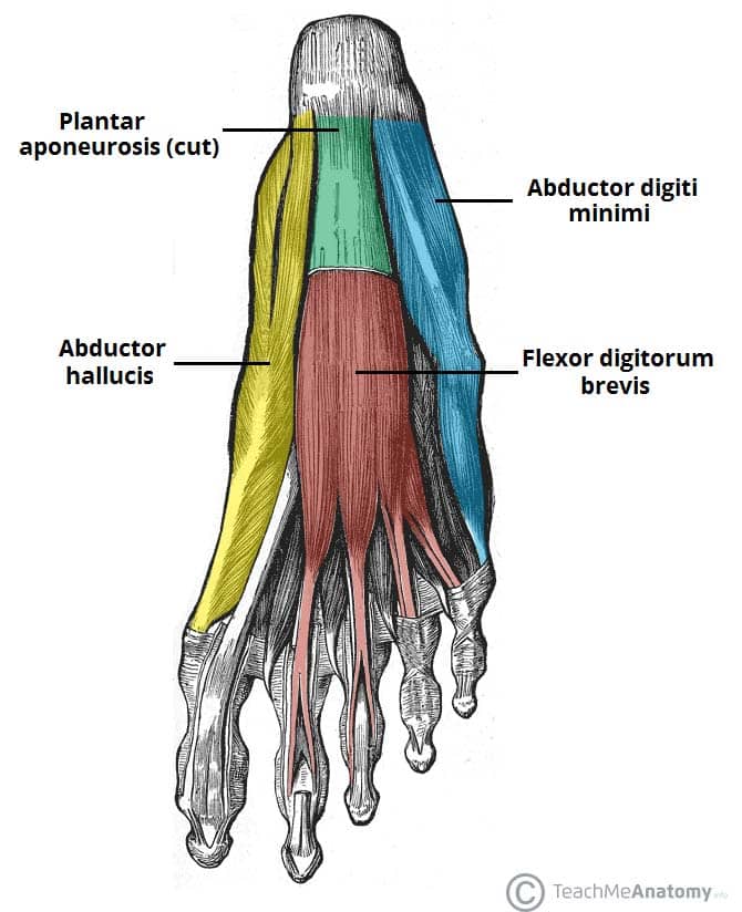
Fig 2 – The first layer of plantar muscles. The plantar aponeurosis has been cut to reveal the underlying flexor digitorum.
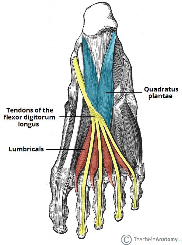
Second Layer
The second plantar layer contains two muscles – the quadratus plantae and the lumbricals. The tendons of the flexor digitorum longus (an extrinsic muscle) also travel through this layer.
Quadratus Plantae
The quadratus plantae is a flat, square-shaped muscle with two heads of origin.
- Attachments: Originates from the medial and lateral plantar surface of the calcaneus. It attaches to the tendons of flexor digitorum longus.
- Actions: Assists the flexor digitorum longus in flexion of the lateral four toes.
- Innervation: Lateral plantar nerve.
Lumbricals
There are four lumbrical muscles in the foot. They are each located medial to their respective tendon of the flexor digitorum longus.
- Attachments: Originates from the tendons of flexor digitorum longus. Attaches to the extensor hoods of the lateral four digits.
- Actions: Flexion at the metatarsophalangeal joints and extension at the interphalangeal joints.
- Innervation:
- Medial lumbrical – medial plantar nerve.
- Lateral three lumbricals – lateral plantar nerve
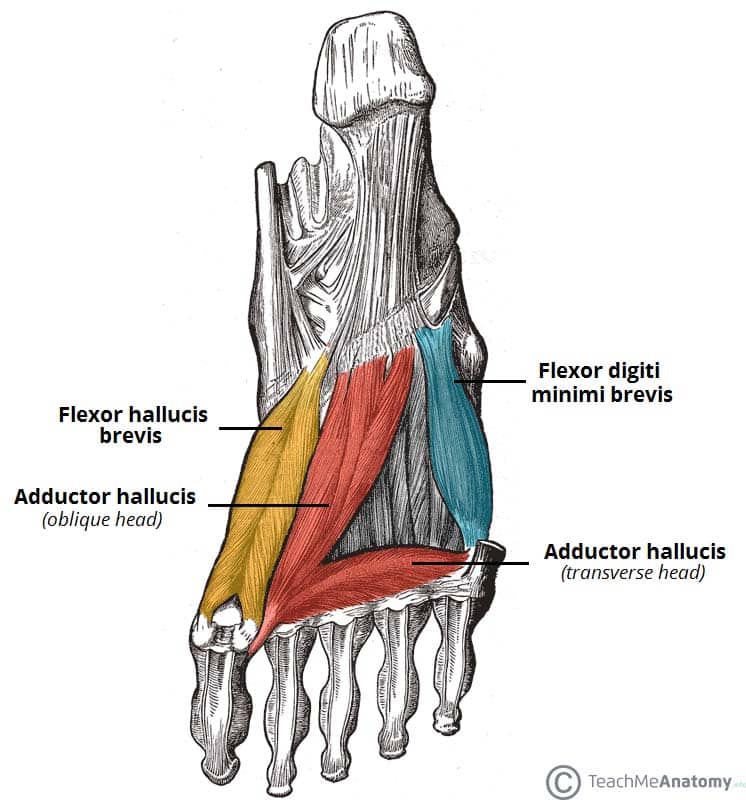
Third Layer
The third layer contains three muscles. The flexor hallucis brevis and adductor hallucis are associated with movements of the great toe. The remaining muscle, the flexor digiti minimi brevis, moves the little toe.
Flexor Hallucis Brevis
The flexor hallucis brevis muscle is located on the medial side of the foot. It has two heads of origin.
- Attachments:
- Lateral head – originates from the plantar surfaces of the cuboid and lateral cuneiforms
- Medial head – originates from the tendon of the posterior tibialis tendon.
- The fibres converge into a single muscle belly, which attaches to the base of the proximal phalanx of the great toe.
- Actions: Flexion of the great toe at the metatarsophalangeal joint.
- Innervation: Medial plantar nerve.
Adductor Hallucis
The adductor hallucis muscle is located laterally to the flexor hallucis brevis. It consists of an oblique and transverse head.
- Attachments:
- Oblique head – originates from the bases of the 2nd, 3rd, and 4th metatarsals.
- Transverse head – originates from the plantar ligaments of the metatarsophalangeal joints.
- Both heads attach to the lateral aspect of the base of the proximal phalanx of the great toe.
- Actions: Adduction of the great toe. Supports the transverse arch of the foot.
- Innervation: Deep branch of lateral plantar nerve.
Flexor Digiti Minimi Brevis
The flexor digiti minimi brevis muscle is located on the lateral side of the foot, underneath the metatarsal of the little toe. It resembles the interossei in structure.
- Attachments: Originates from the base of the fifth metatarsal and attaches to the base of the proximal phalanx of the fifth digit.
- Actions: Flexion of the little toe at the metatarsophalangeal joint.
- Innervation: Superficial branch of lateral plantar nerve.
Fourth Layer
The plantar and dorsal interossei comprise the fourth and final plantar muscle layer. The plantar interossei have a unipennate shape, while the dorsal interossei are bipennate.
Plantar Interossei
There are three plantar interossei, which are located between the metatarsals. Each arises from a single metatarsal.
- Attachments: Originates from the medial side of metatarsals three to five. Attaches to the medial sides of the phalanges of digits three to five.
- Actions: Adduction of the lateral three digits and flexion at the metatarsophalangeal joints.
- Innervation: Lateral plantar nerve.
Dorsal Interossei
There are four dorsal interossei, which are located between the metatarsals. Each arises from two metatarsals.
- Attachments: Originates from the lateral aspect of the metatarsals. The first muscle attaches to the medial side of the proximal phalanx of the second digit. The second to fourth interossei attach to the lateral sides of the proximal phalanxes of digits two to four.
- Actions: Abduction of the lateral four digits and flexion at the metatarsophalangeal joints.
- Innervation: Lateral plantar nerve.
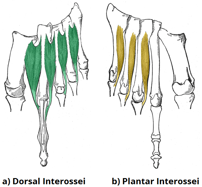
Fig 5 – The fourth layer of the plantar muscles. Note the unipennate shape of the plantar interossei, and the bipennate shape of the dorsal interossei
Clinical Relevance: Medial Plantar Nerve Entrapment
The medial plantar nerve can become compressed and irritated as it passes deep to the abductor hallucis muscle.
This can cause aching, numbness and paraesthesia on the medial side of the sole of the foot. The muscle can become compressed during repetitive eversion of the foot, which may occur in some sports such as gymnastics.