The foot has three arches: two longitudinal (medial and lateral) arches and one anterior transverse arch (Fig. 1). They are formed by the tarsal and metatarsal bones, and supported by ligaments and tendons in the foot.
Their shape allows them to act in the same way as a spring, bearing the weight of the body and absorbing the shock produced during locomotion. The flexibility conferred to the foot by these arches facilitates functions such as walking and running.
In this article, we shall examine the anatomy of the arches of the foot – their bony and ligamentous structure, the supporting tendons, and their clinical correlations.
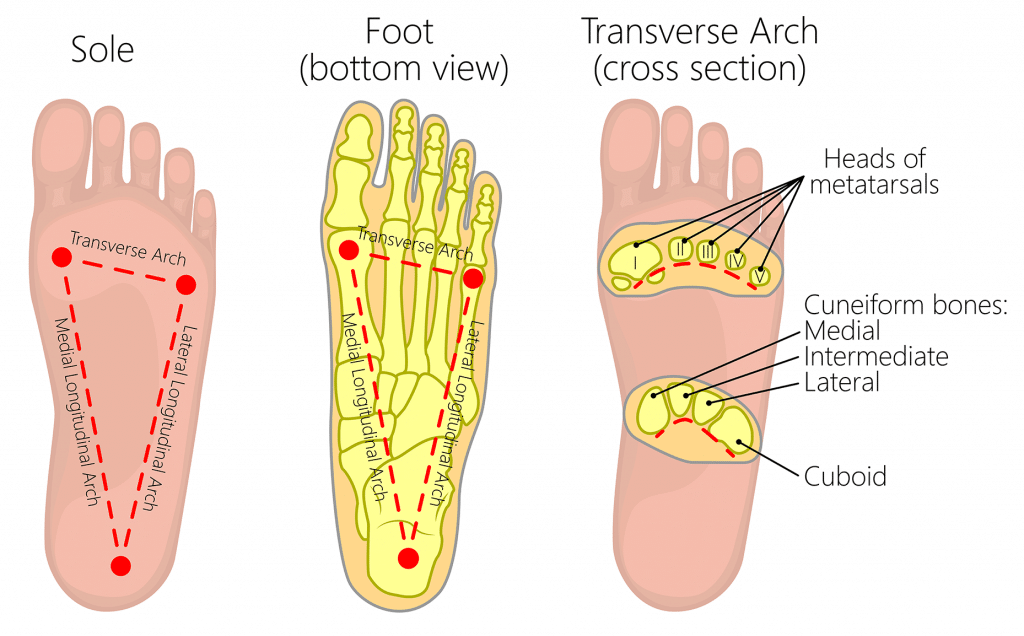
Fig 1 – The transverse, medial longitudinal and lateral longitudinal arches of the foot.
Longitudinal Arches
There are two longitudinal arches in the foot – the medial and lateral arches. They are formed between the tarsal bones and the metatarsal heads
Medial Arch
The medial arch is the higher of the two longitudinal arches. It is formed by the calcaneus, talus, navicular, three cuneiforms and first three metatarsal bones. It is supported by:
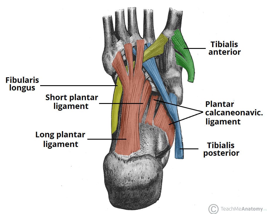
- Muscular support: Tibialis anterior and posterior, fibularis longus, flexor digitorum longus, flexor hallucis, and the intrinsic foot muscles
- Ligamentous support: Plantar ligaments (in particular the long plantar, short plantar and plantar calcaneonavicular ligaments), medial ligament of the ankle joint.
- Bony support: Shape of the bones of the arch.
- Other: Plantar aponeurosis.
Lateral Arch
The lateral arch is the flatter of the two longitudinal arches, and lies on the ground in the standing position. It is formed by the calcaneus, cuboid and 4th and 5th metatarsal bones. It is supported by:
- Muscular support: Fibularis longus, flexor digitorum longus, and the intrinsic foot muscles.
- Ligamentous support: Plantar ligaments (in particular the long plantar, short plantar and plantar calcaneonavicular ligaments).
- Bony support: Shape of the bones of the arch.
- Other: Plantar aponeurosis.
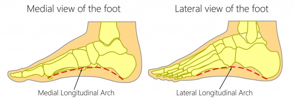
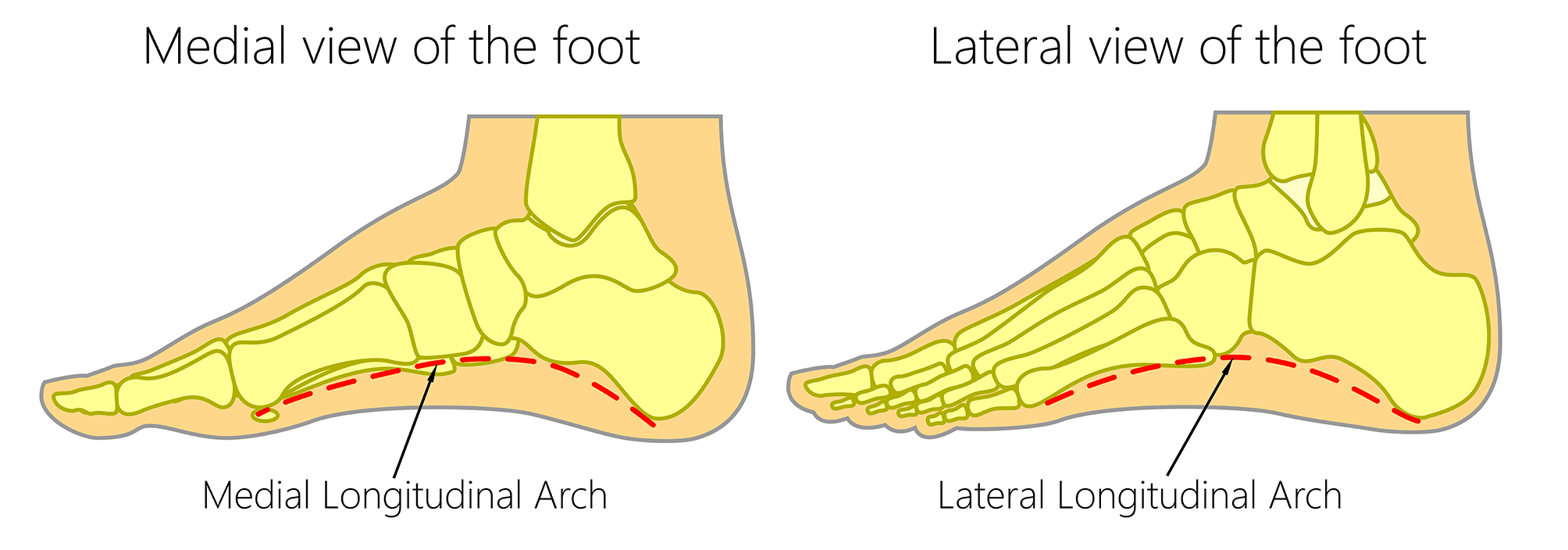
Fig 2 – The medial and lateral longitudinal arches of the foot.
Transverse Arch
The transverse arch is located in the coronal plane of the foot. It is formed by the metatarsal bases, the cuboid and the three cuneiform bones. It has:
- Muscular support: Fibularis longus and tibialis posterior.
- Ligamentous support: Plantar ligaments (in particular the long plantar, short plantar and plantar calcaneonavicular ligaments) and deep transverse metatarsal ligaments.
- Other support: Plantar aponeurosis.
- Bony support: The wedged shape of the bones of the arch.
Clinical Relevance – Pes Cavus (High Arches)
Pes cavus is a foot condition characterised by an unusually high medial longitudinal arch. It can appear in early life and become symptomatic with increasing age. Due to the higher arch, the ability to shock absorb during walking is diminished and an increased degree of stress is placed on the ball and heel of the foot.
Consequently, symptoms will generally include pain in the foot, which can radiate to the ankle, leg, thigh and hip. This pain is transmitted up the lower limb from the foot due to the unusually high stress placed on the hindfoot during the heel strike of the gait cycle.
Causes of pes cavus can be idiopathic, hereditary, due to an underlying congenital foot problem such as club foot, or secondary to neuromuscular damage such as in poliomyelitis.
The condition is generally managed by supporting the foot through the use of special shoes or sole cushioning inserts. Reducing the amount of weight the foot has to bear, via overall weight loss can also improve the symptoms.
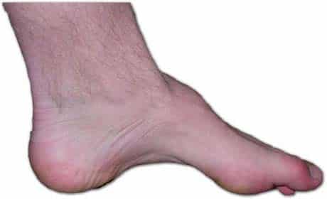
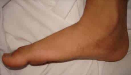
Fig 4 – Pes cavus, an abnormally high longitudinal arch.
Clinical Relevance: Pes Planus (Flat Footed)
Pes planus is a common condition in which the longitudinal arches have been lost. Arches do not develop until about 2-3 years of age, meaning flat feet during infancy is normal.
Because the arches are formed, in part, by the tight tendons of the foot, damage to these tissues through direct injury or trauma can cause pes planus. However in some people, the arches never formed during development.
For most individuals, being flat-footed causes few, if any, symptoms. In children it may result in foot and ankle pain, whereas in adults the feet may ache after prolonged activity.
Treatment, if indicated, generally involves the use of arch-supporting inserts for shoes.