The popliteal fossa is a diamond shaped area located on the posterior aspect of the knee. It is the main path by which vessels and nerves pass between the thigh and the leg.
In this article, we shall look at the anatomy of the popliteal fossa – its borders, contents and clinical correlations.
Borders
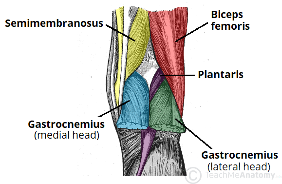
The popliteal fossa is diamond shaped with four borders. These borders are formed by the muscles in the posterior compartment of the leg and thigh:
- Superomedial – semimembranosus.
- Superolateral – biceps femoris.
- Inferomedial – medial head of the gastrocnemius.
- Inferolateral – lateral head of the gastrocnemius and plantaris.
The floor of the popliteal fossa is formed by the posterior surface of the knee joint capsule, popliteus muscle and posterior femur. The roof is made of up two layers: popliteal fascia and skin. The popliteal fascia is continuous with the fascia lata of the leg.

Fig 1 – The borders of the popliteal fossa are formed by the muscles of the thigh and leg.
Contents
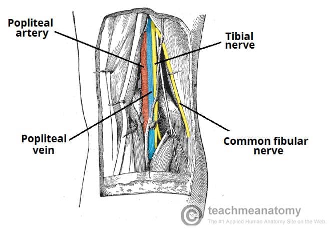
The popliteal fossa is the main conduit for neurovascular structures entering and leaving the leg. Its contents are (medial to lateral):
- Popliteal artery
- Popliteal vein
- Tibial nerve
- Common fibular nerve (common peroneal nerve)
The tibial and common fibular nerves are the most superficial of the contents of the popliteal fossa. They are both branches of the sciatic nerve. The common fibular nerve follows the biceps femoris tendon, travelling along the lateral margin of the popliteal fossa.
The small saphenous vein pierces the popliteal fascia and passes between the two heads of gastrocnemius to empty into the popliteal vein.
In the popliteal fossa, the deepest structure is the popliteal artery. It is a continuation of the femoral artery, and travels into the leg to supply it with blood.
Clinical Relevance: Swelling in the Popliteal Fossa
The appearance of a mass in the popliteal fossa has many differential diagnoses. The two main causes are baker’s cyst and aneurysm of the popliteal artery.
Baker’s Cyst
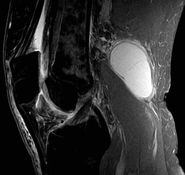
A Baker’s cyst (popliteal cyst) refers to the inflammation and swelling of the semimembranosus bursa – a sac-like structure containing a small amount of synovial fluid. It usually arises in conjunction with osteoarthritis of the knee.
Whilst it usually self-resolves, the cyst can rupture and produce symptoms similar to deep vein thrombosis.
Popliteal Aneurysm
An aneurysm is a dilation of an artery, which is greater than 50% of the normal diameter. The popliteal fascia (the roof of the popliteal fossa) is tough and non-extensible, and so an aneurysm of the popliteal artery has consequences for the other contents of the popliteal fossa.
The tibial nerve is particularly susceptible to compression from the popliteal artery. The major features of tibial nerve compression are:
- Weakened or absent plantar flexion
- Paraesthesia of the foot and posterolateral leg
An aneurysm of the popliteal artery can be detected by an obvious palpable pulsation in the popliteal fossa. An arterial bruit may be heard on auscultation.
Other Causes
Rarer causes of a popliteal mass include deep vein thrombosis, adventitial cyst of the popliteal artery and various neoplasms (such as rhabdomyosarcoma).

