The nasal skeleton is a combination of bone and cartilage which forms both what we can see as the external nose and the internal nasal septum – which divides the two nasal cavities of the head.
Here we will discuss the anatomy of the nasal skeleton and its component bones.
Anatomical Structure
The skeleton of the nose is formed by three types of tissue; bone, cartilage and fibro-fatty tissue. When looking at the scaffolding of the nose, it is useful to divide the structures into two parts; the external nasal skeleton and internal nasal septum.
External Nasal Skeleton
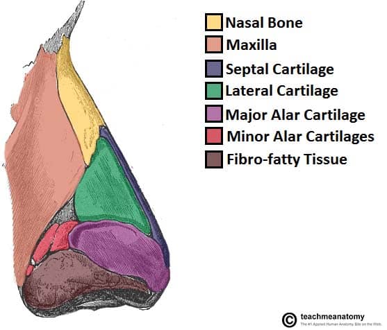
The external skeleton extends the nasal cavities onto the front of the face (see Figure 1). It is partly formed by the nasal and maxillary bones, which are situated superiorly.
The inferior portion of the external nose is made up of hyaline cartilages; lateral, major alar, minor alar, and the cartilaginous septum. The lateral and major alar cartilages are the largest, and contribute the most to the shape of the nose here. The minor alar cartilages vary in number, there are usually 3 or 4 on each side.
Internal Nasal Septum
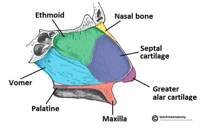
The internal nasal septum separates the nasal cavity into two nostrils. The bones that contribute to the nasal septum can be divided into:
- Paired bones: Nasal, maxillary and palatine bones
- Unpaired bones: Ethmoid and vomer bones.
In addition to the bones of the nose, the septal and greater alar cartilages also constitute part of the nasal septum.
The ethmoid contributes to the central portion of nasal septum. It is one of the most complex bones in the human body, and its structure is beyond the scope of this article, however more information can be found here. The anterior and posterior parts are formed by the septal cartilage and vomer bone respectively.
The floor of the nasal cavity is formed by the hard palate, separating it from the oral cavity. The hard palate consists of the palatine bone posteriorly, and the palatine process of the maxilla anteriorly. The cribriform plate of the ethmoid bone forms the roof of the nasal cavity.
Clinical Relevance: Nasal Fracture
Due to the prominence of the external nasal skeleton, nasal fractures are common – the most common facial fracture.
Fractures usually occur as a result of blunt trauma to the nose. A common sequela of nasal fractures is permanent deformity, due to disruption of the bone and cartilage.

