The muscles of mastication are associated with movements of the jaw (temporomandibular joint). There are four muscles:
- Masseter
- Temporalis
- Medial pterygoid
- Lateral pterygoid
The muscles of mastication develop from the first pharyngeal arch. They are therefore innervated by a branch of the trigeminal nerve (CN V), the mandibular nerve.
In this article, we shall look at the anatomy of the muscles of mastication – their attachments, actions, and innervation.
(NB: It is important to note that all the muscles mentioned here are bilateral structures).
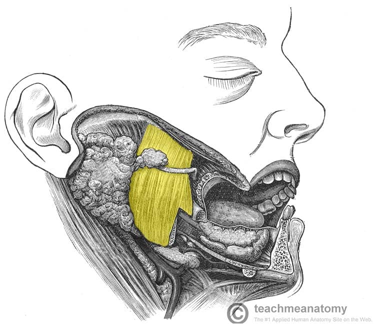
Masseter
The masseter muscle is the most powerful muscle of mastication. It is quadrangular in shape and has two parts: deep and superficial.
It lies superficial to the pterygoids and temporalis muscles.
- Attachments:
- The superficial part originates from maxillary process of the zygomatic bone.
- The deep part originates from the zygomatic arch of the temporal bone.
- Both parts attach to the ramus of the mandible.
- Actions: Elevation of the mandible (closes the mouth).
- Innervation: Mandibular nerve (V3).
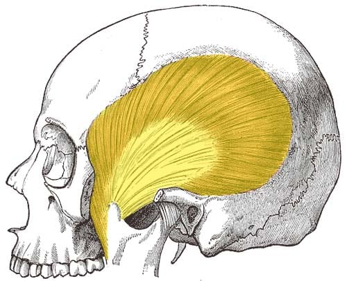
Temporalis
The temporalis muscle originates from the temporal fossa – a shallow depression on the lateral aspect of the skull.
The muscle is covered by tough fascia which can be harvested surgically and used to repair a perforated tympanic membrane (an operation known as a myringoplasty).
- Attachments: Originates from the temporal fossa of the skull and attaches onto the coronoid process of the mandible.
- Actions: Elevation of the mandible (closing the mouth). Also performs retraction of the mandible (moving the jaw posteriorly).
- Innervation: Mandibular nerve (V3).
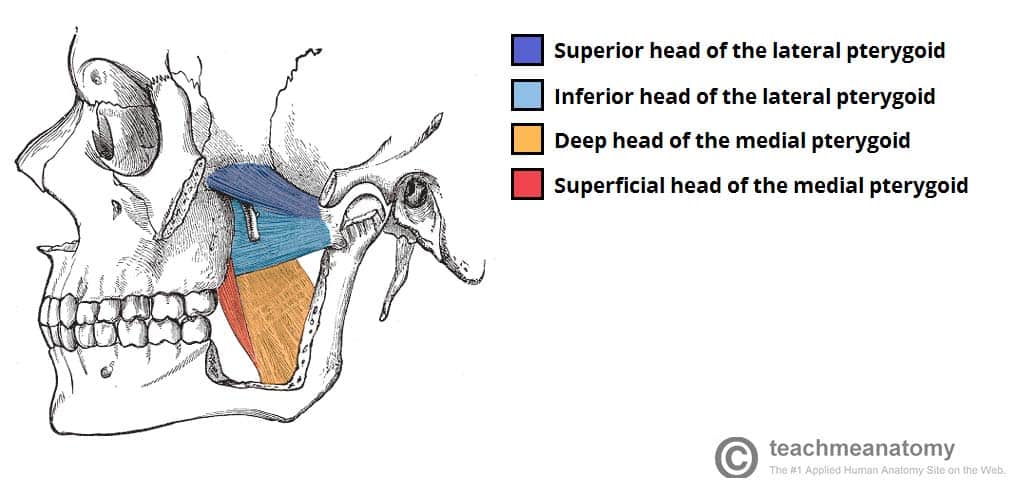
Medial Pterygoid
The medial pterygoid muscle has a quadrangular shape with two heads: deep and superficial. It is located inferiorly to the lateral pterygoid.
- Attachments:
- The superficial head originates from the maxillary tuberosity and the pyramidal process of palatine bone.
- The deep head originates from the medial aspect of the lateral pterygoid plate of the sphenoid bone.
- Both heads attach to the ramus of the mandible near the angle of mandible.
- Actions: Elevation of the mandible (closing the mouth).
- Innervation: Mandibular nerve (V3).
Lateral Pterygoid
The lateral pterygoid muscle has a triangular shape with two heads: superior and inferior. It has horizontally orientated muscle fibres, and thus is the major protractor of the mandible.
- Attachments:
- The superior head originates from the greater wing of the sphenoid.
- The inferior head originates from the lateral pterygoid plate of the sphenoid.
- The two heads converge into a tendon which attaches to the neck of the mandible.
- Actions:
- Bilateral action – protraction of the mandible and depression of the chin.
- Unilateral action – ‘side to side’ movement of the jaw.
- Innervation: Mandibular nerve (V3).