The floor of the cranial cavity is divided into three distinct depressions. They are known as the anterior cranial fossa, middle cranial fossa and posterior cranial fossa. Each fossa accommodates a different part of the brain.
The middle cranial fossa is located, as its name suggests, centrally in the cranial floor. It is said to be “butterfly shaped”, with a middle part accommodating the pituitary gland and two lateral parts accommodating the temporal lobes of the brain
In this article, we shall look at the borders, contents and clinical correlations of the middle cranial fossa.
Borders
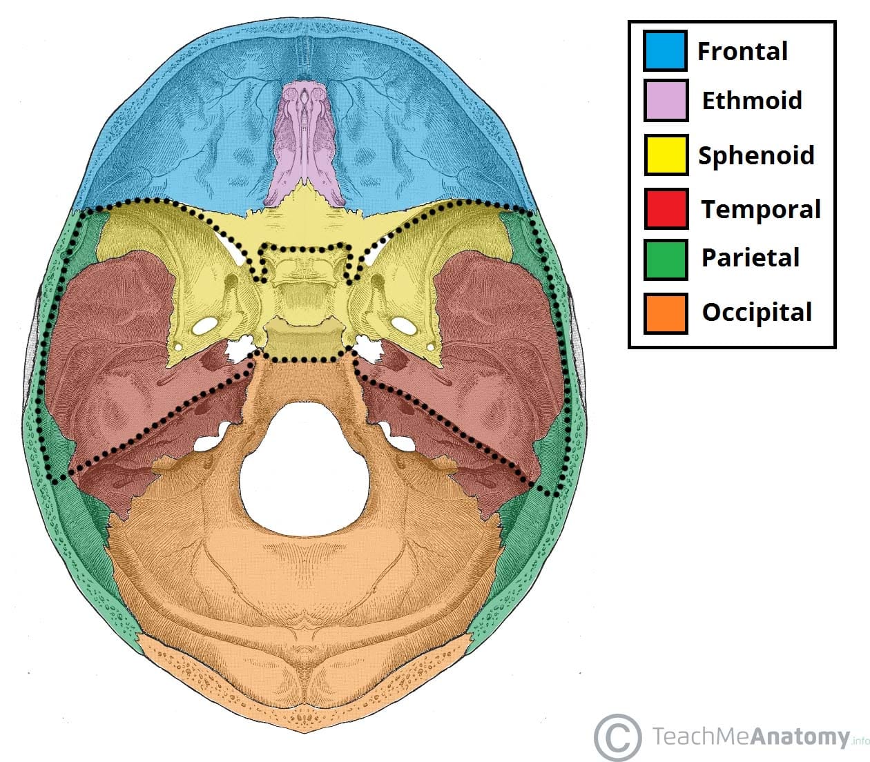
The middle cranial fossa consists of three bones – the sphenoid bone and the two temporal bones.
Its boundaries are as follows:
- Anteriorly and laterally it is bounded by the lesser wings of the sphenoid bone. These are two triangular projections of bone that arise from the central sphenoid body.
- Anteriorly and medially it is bounded by the limbus of the sphenoid bone. The limbus is a bony ridge that forms the anterior border of the chiasmatic sulcus (a groove running between the right and left optic canals).
- Posteriorly and laterally it is bounded by the superior border of the petrous part of the temporal bone.
- Posteriorly and medially it is bounded by the dorsum sellae of the sphenoid bone. This is a large superior projection of bone that arises from the sphenoidal body.
- The floor is formed by the body and greater wing of the sphenoid, and the squamous and petrous parts of the temporal bone.

Fig 1.0 – The bones of the cranial floor. The middle cranial fossa has been highlighted.
Contents
The middle cranial fossa consists of a central portion, which contains the pituitary gland, and two lateral portions, which accommodate the temporal lobes of the brain.
Both parts of the fossa are marked by numerous bony landmarks, which will be discussed below.
Central Part
The central part of the middle cranial fossa is formed by the body of the sphenoid bone. It contains the sella turcica (latin for Turkish saddle), which is a saddle-shaped bony prominence (see Fig 1.2). It acts to hold and support the pituitary gland, and consists of three parts:
- The tuberculum sellae (horn of the saddle) is a vertical elevation of bone. It forms the anterior wall of the sella turcica, and the posterior aspect of the chiasmatic sulcus (a groove running between the right and left optic canals).
- The hypophysial fossa or pituitary fossa (seat of the saddle) sits in the middle of the sella turcica. It is a depression in the body of the sphenoid, which holds the pituitary gland.
- The dorsum sellae (back of the saddle) forms the posterior wall of the sella turcica. It is a large square of bone, pointing upwards and forwards. It separates the middle cranial fossa from the posterior cranial fossa.
The sella turcica is surrounded by the anterior and posterior clinoid processes. The anterior clinoid processes arise from the sphenoidal lesser wings, while the posterior clinoid processes are the superolateral projections of the dorsum sellae. They serve as attachment points for the tentorium cerebelli, a membranous sheet that divides the brain.
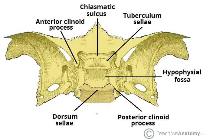
Fig 1.1 – Bony landmarks of the central part of the middle cranial fossa.
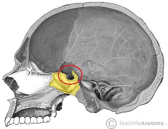
Fig 1.2 – Sagittal section of the skull, showing the saddle-like sella turcica.
Lateral Parts
The depressed lateral parts of the middle cranial fossa are formed by the greater wings of the sphenoid bone, and the squamous and petrous parts of the temporal bones. They support the temporal lobes of the brain. It is the site of many foramina – small holes by which vessels and nerves enter and leave the cranial cavity.
Foramina
There are many foramina that transmit vessels and nerves into and out of the middle cranial fossa. These foramina will be discussed in relation to the bones they are situated in.
Foramina of the Sphenoid Bone
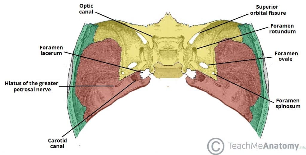
The optic canals are situated anteriorly in the middle cranial fossa. They transmit the optic nerves (CN II) and ophthalmic arteries into the orbital cavities. The optic canals are connected by the chiasmatic sulcus, a depressed groove running transversely between the two.
Immediately lateral to the central part of the middle cranial fossa are four foramina:
- The superior orbital fissure opens anteriorly into the orbit. It transmits the oculomotor nerve (CN III), trochlear nerve (CN IV), ophthalmic branch of the trigeminal nerve (CN V1), abducens nerve (CN VI), ophthalmic veins and sympathetic fibres.
- The foramen rotundum opens into the pterygopalatine fossa and transmits the maxillary branch of the trigeminal nerve (CN V2).
- The foramen ovale opens into the infratemporal fossa, transmitting the mandibular branch of the trigeminal nerve (CN V3) and accessory meningeal artery.
- The foramen spinosum also opens into the infratemporal fossa. It transmits the middle meningeal artery, middle meningeal vein and a meningeal branch of CN V3.
Foramina of the Temporal Bone
The temporal bone is marked by three major foramina:
- Hiatus of the greater petrosal nerve – transmits the greater petrosal nerve (a branch of the facial nerve), and the petrosal branch of the middle meningeal artery.
- Hiatus of the lesser petrosal nerve – transmits the lesser petrosal nerve (a branch of the glossopharyngeal nerve).
- Carotid canal – located posteriorly and medially to the foramen ovale. This is traversed by the internal carotid artery, which ascends into the cranium to supply the brain with blood. The deep petrosal nerve also passes through this canal.
At the junction of the sphenoid, temporal and occipital bones is the foramen lacerum. In life, this foramen is filled with cartilage, which is pierced only by small blood vessels.
Clinical Relevance: Pituitary Surgery
The pituitary gland lies in the sella turcica of the sphenoid bone, within the middle cranial fossa. In cases of a pituitary tumour, it may need to be removed surgically.
Surgery to remove pituitary tumours is usually by a endoscopic transsphenoidal approach. An endoscope is inserted through the nostrils, or more rarely through an incision into either the upper lip or nasal septum. It is then advanced through the nasal cavity. The sphenoid sinus is opened and the endoscope passes through to the pituitary gland where it lies on the sella turcica. The tumour can then be removed in sections.
Complications of pituitary surgery include CSF rhinorrhoea, meningitis, diabetes insipidus, haemorrhage and visual disturbances.