The superior mesenteric artery (SMA) is a major artery of the abdomen.
It arises from the abdominal aorta, and supplies arterial blood to the organs of the midgut – which spans from the major duodenal papilla (of the duodenum) to the proximal 2/3 of the transverse colon.
In this article, we shall look the anatomy of the superior mesenteric artery – its anatomical position, branches, anastomoses, and clinical relevance.
Anatomical Position
The superior mesenteric artery is the second of the three major anterior branches of the abdominal aorta (the other two are the coeliac trunk and inferior mesenteric artery).
It arises anteriorly from the abdominal aorta at the level of the L1 vertebrae, immediately inferior to the origin of the coeliac trunk.
After arising from the abdominal aorta, the superior mesenteric artery descends down the posterior aspect of the abdomen. At this point, it has several important anatomical relations:
- Anterior – pyloric part of the stomach, splenic vein and neck of the pancreas.
- Posterior – left renal vein, uncinate process of the pancreas and inferior part of the duodenum.
- The uncinate process hooks around the back of the superior mesenteric artery
Major Branches
The superior mesenteric artery gives rise to various branches that supply the small intestines, cecum, ascending and part of the transverse colon (Fig 1).
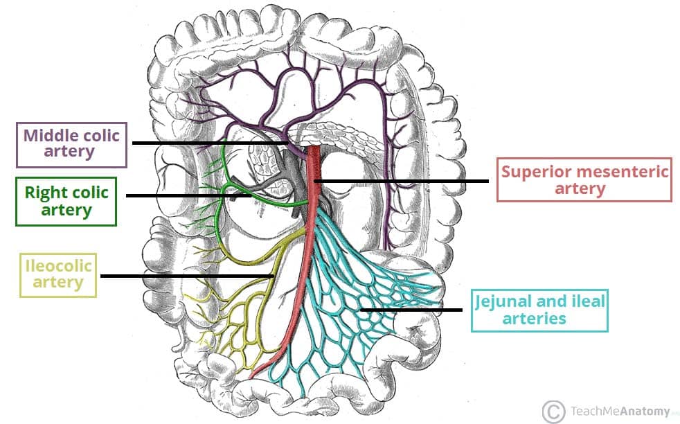
Fig 1 – The superior mesenteric artery and its branches. Note: the inferior pancreatoduodenal artery arises more proximally, and is not visible on this illustration.
Inferior Pancreaticoduodenal Artery
The inferior pancreaticoduodenal artery is the first branch of the SMA. It forms anterior and posterior vessels, which anastomose with branches of the superior pancreaticoduodenal artery (derived from the coeliac trunk). This network supplies the inferior region of the head of the pancreas, the uncinate process, and the duodenum.
Jejunal and Ileal Arteries
The superior mesenteric artery gives rise to numerous arteries that supply the jejunum and ileum.
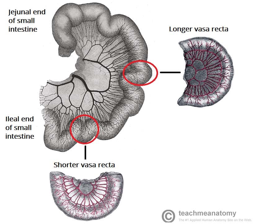
The arteries pass between the layers of the mesentery and form anastomotic arcades – from which smaller, straight arteries (known as the “vasa recta”) arise to supply the organs (fig 2).
The jejunal blood supply is characterised by a smaller number of arterial arcades, but longer vasa recta. In contrast, the ileal blood supply is marked by more arterial arcades with shorter vasa recta.
Middle and Right Colic Arteries
The right and middle colic arteries arise from the right side of the superior mesenteric artery to supply the colon:
- Middle colic artery – supplies the transverse colon.
- Right colic artery – supplies the ascending colon.
Ileocolic Artery
The ileocolic artery is the final major branch of the superior mesenteric artery. It passes inferiorly and to the right, giving rise to branches to the ascending colon, appendix, cecum, and ileum. In cases of appendectomy, the appendicular artery is ligated.
Clinical Relevance: Occlusion of the Superior Mesenteric Artery
There are a number of cause of superior mesenteric artery occlusion, including thrombosis, embolism, abdominal aortic aneurysm and aortic dissection.
Often acute, occlusion of the SMA restricts blood flow to the midgut, resulting in intestinal ischaemia. It is more common in the elderly, and most usually presents with abdominal pain. The most useful investigation in this scenario is CT scan of the abdomen.
Treatment is surgical.