There are two venous systems that drain abdominal structures – the portal venous system and the systemic venous system. The portal system transports venous blood to the liver for processing, whilst the systemic venous system returns blood to the right atrium of the heart.
In this article, we shall consider the anatomy of these two venous systems – the major vessels involved, their anatomical course, and their tributaries.
Premium Feature
3D Model
Systemic Venous System
The systemic venous system transports deoxygenated blood to the right atrium of the heart. The major vessel in this system is the inferior vena cava.
Inferior Vena Cava
The inferior vena cava is the common convergence of venous drainage from all structures below the diaphragm. It is located on the posterior abdominal wall; anteriorly to the vertebral column and to the right of the abdominal aorta.
The vessel is formed by the union of the common iliac veins at the L5 vertebral level. It ascends superiorly, and leaves the abdomen by piercing the central tendon of the diaphragm at the T8 level (the caval hiatus). Within the thorax, the inferior vena cava drains into the right atrium of the heart.
During its long course, the inferior vena cava shares an anatomical relationship with numerous abdominal structures – including the right common iliac artery, the root of the mesentery, the head of the pancreas, the bile duct, the portal vein and the liver.
Tributaries
The inferior vena cava is responsible for the venous drainage of all structures below the diaphragm. It receives tributaries from:
- Common iliac veins – formed by the external and internal iliac veins. They drain the lower limbs and gluteal region.
- Lumbar veins – drain the posterior abdominal wall.
- Renal veins – drain the kidneys, left adrenal gland and left testis/ovary.
- Right testicular or ovarian vein – drains the right testes in males and the right ovary in females (the left testicular or ovarian vein drains into the left renal vein).
- Right suprarenal vein – drains the right adrenal gland (the left adrenal vein drains into the left renal vein).
- Inferior phrenic veins – drain the diaphragm.
- Hepatic veins – drain the liver.
There are no tributaries from the spleen, pancreas, gallbladder or the abdominal part of the GI tract – as these structures are first drained into the portal venous system. However, venous return from these structures ultimately enters the inferior vena cava via the hepatic veins (after being processed by the liver).
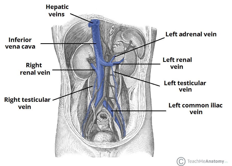
Fig 1.0
The inferior vena cava and major tributaries. Note how the left adrenal vein and left testicular vein empty into the left renal vein.
Premium Feature
Dissection Images

Portal Venous System
The portal system carries venous blood (rich in nutrients that have been extracted from food) to the liver for processing.
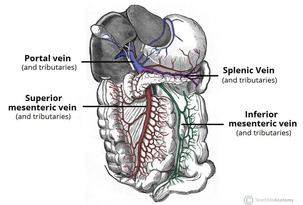
The major vessel of the portal system is the portal vein. It is the point of convergence for the venous drainage of the spleen, pancreas, gallbladder and the abdominal part of the gastrointestinal tract. The portal vein is formed by the union of the splenic vein and the superior mesenteric vein, posterior to the neck of the pancreas, at the level of L2.
As it ascends towards the liver, the portal vein passes posteriorly to the superior part of the duodenum and the bile duct. Immediately before entering the liver, the portal vein divides into right and left branches which then enter the parenchyma of the liver separately.
Tributaries
The portal vein is formed by the union of the splenic vein and superior mesenteric vein. It receives additional tributaries from:
- Right and left gastric veins – drain the stomach.
- Cystic veins – drains the gallbladder.
- Para-umbilical veins – drain the skin of the umbilical region.
Splenic Vein
The splenic vein is formed from a variety of smaller vessels as they leave the hilum of the spleen.
Unlike the splenic artery, the splenic vein is straight and it maintains contact with the body of the pancreas as it crosses the posterior abdominal wall. As it reaches the neck of the pancreas, the splenic vein joins the superior mesenteric vein to form the portal vein.
Tributaries
Tributaries to the splenic vein include:
- Short gastric veins – drain the fundus of the stomach.
- Left gastro-omental vein – drains the greater curvature of the stomach.
- Pancreatic veins – drain the pancreas.
- Inferior mesenteric vein – drains the colon.
The inferior mesenteric vein drains blood from the rectum, sigmoid colon, descending colon and splenic flexure. It begins as the superior rectal vein and ascends, receiving tributaries from the sigmoid veins and the left colic veins. As it ascends further it passes posteriorly to the body of the pancreas and typically joins the splenic vein.
Superior Mesenteric Vein
The superior mesenteric vein drains blood from the small intestine, cecum, ascending colon and transverse colon. It begins in the right iliac fossa, as a convergence of the veins draining the terminal ileum, cecum and appendix. It ascends within the mesentery of the small intestine, and then travels posteriorly to the neck of the pancreas to join the splenic vein.
Tributaries
Tributaries to the superior mesenteric vein include:
- Right gastro-omental vein – drains the greater curvature of the stomach.
- Anterior and posterior inferior pancreaticoduodenal veins – drain the pancreas and duodenum.
- Jejunal vein – drain the jejunum.
- Ileal vein – drain the ileum.
- Ileocolic vein – drains the ileum, colon and cecum.
- Right colic vein – drains the ascending colon.
- Middle colic vein – drains the transverse colon.
Many of these tributaries are formed as an accompanying vein for each branch of the superior mesenteric artery.
Clinical Relevance – Porto-Systemic Anastomoses
A porto-systemic anastomosis is a connection between the veins of the portal venous system, and the veins of the systemic venous system. The major sites of these anastomoses include:
- Oesophageal – Between the oesophageal branch of the left gastric vein and the oesophageal tributaries to the azygous system.
- Rectal – Between the superior rectal vein and the inferior rectal veins.
- Retroperitoneal – Between the portal tributaries of the mesenteric veins and the retroperitoneal veins.
- Paraumbilical – Between the portal veins of the liver and the veins of the anterior abdominal wall.

Fig 3
Endoscopic appearance of oesophageal varices. They can undergo rupture, leading to large volumes of blood loss.
When blood flow through the portal system is obstructed (e.g due to cirrhosis, portal vein thrombosis, or external pressure from a tumour), the pressure within portal system increases. A portal venous pressure in excess of 20mmHg is defined as portal hypertension.
In portal hypertension, blood may be re-directed through the porto-systemic anastomoses (as these are now under a lower pressure). If a large volume of blood passes through these anastomoses over a long period of time, the veins around the anastomosis can become abnormally dilated – known as varices. Rupture of oesophageal or rectal varices can result in fatal blood loss.