The larynx (voice box) is an organ located in the anterior neck. It is a component of the respiratory tract, and has several important functions, including phonation, the cough reflex, and protection of the lower respiratory tract.
The muscles of the larynx can be divided into two groups; the external muscles and the internal muscles. The external muscles act to elevate or depress the larynx during swallowing. In contrast, the internal muscles act to move the individual components of the larynx – playing a vital role in breathing and phonation.
In this article, we shall look at the anatomy of the laryngeal muscles – their attachments, innervation and blood supply.
Extrinsic Muscles
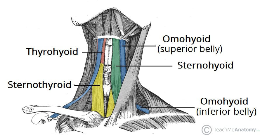
The extrinsic muscles act to move the larynx superiorly and inferiorly. They are comprised of the suprahyoid and infrahyoid groups, and the stylopharyngeus (a muscle of the pharynx).
The supra- and infrahyoid muscle groups attach to the hyoid bone. This, in turn, is bound to the larynx by strong ligaments; allowing the whole of the larynx to be moved as one unit.
As a general rule, the suprahyoid muscles and the stylopharyngeus elevate the larynx, whilst the infrahyoid muscles depress the larynx.
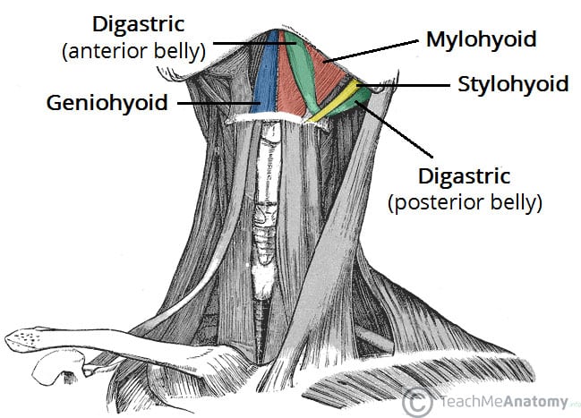
Fig 1 – Anterior view of the neck with the suprahyoid muscles highlighted.
Intrinsic Muscles
The intrinsic laryngeal muscles act on the individual components of the larynx. They control the shape of the rima glottidis (opening between the vocal folds and the arytenoid cartilages), and the length and tension of the vocal folds.
All the intrinsic muscles of the larynx (except the cricothyroid) are innervated by the inferior laryngeal nerve – the terminal branch of the recurrent laryngeal nerve, itself a branch of the vagus nerve. The cricothyroid is innervated by the external branch of the superior laryngeal nerve – again derived from the vagus nerve.
Cricothyroid
The cricothyroid muscle stretches and tenses the vocal ligaments, and so is important for the creation of forceful speech. It also has a role in altering the tone of voice (along with the thyroarytenoid muscle), hence its colloquial name ‘singer’s muscle’.
- Attachments: Originates from the anterolateral aspect of the cricoid cartilage and attaches to the inferior margin and inferior horn of the thyroid cartilage.
- Actions: Stretches and tenses the vocal ligament.
- Innervation: External laryngeal nerve (branch of superior laryngeal).
Thyroarytenoid
The thyroarytenoid muscle acts to relax the vocal ligament, allowing for a softer voice.
- Attachments: Originates from the inferoposterior aspect of the angle of the thyroid cartilage and attaches to the anterolateral part of the arytenoid cartilage.
- Actions: Relaxes the vocal ligament.
- Innervation: Inferior laryngeal nerve (branch of recurrent laryngeal).
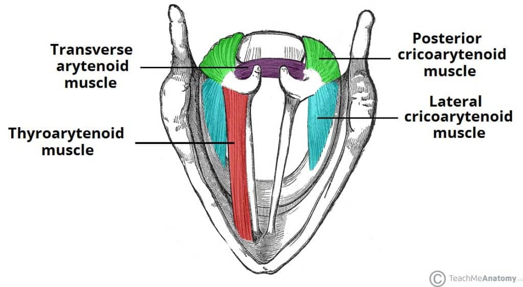
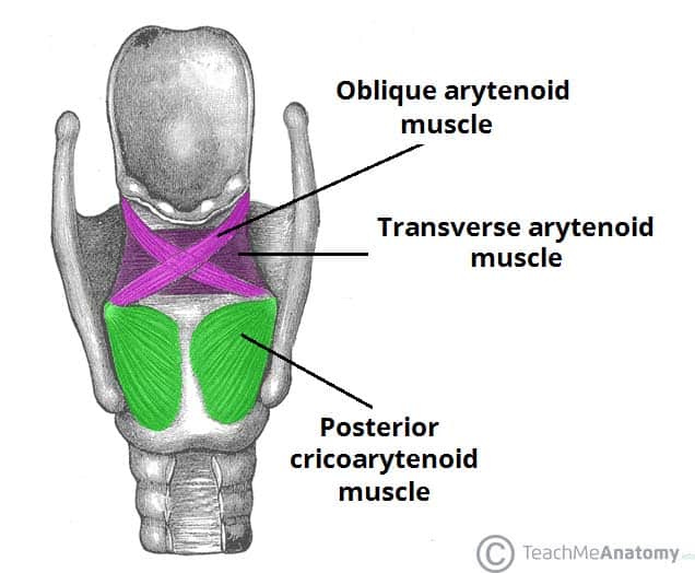
Posterior cricoarytenoid
The posterior cricoarytenoid muscles are the sole abductors of the vocal folds, and thus the only muscle capable of widening the rima glottidis.
- Attachments: Originates from the posterior surface of the cricoid cartilage and attaches to the muscular process of the arytenoid cartilage.
- Actions: Abduction of the vocal folds.
- Innervation: Inferior laryngeal nerve (branch of recurrent laryngeal).
Lateral cricoarytenoid
The lateral cricoarytenoid muscles are the major adductors of the vocal folds. They narrow the rima glottidis, modulating the tone and volume of speech.
- Attachments: Originates from the arch of the cricoid cartilage and attaches to the muscular process of the arytenoid cartilage.
- Actions: Adduction of the vocal folds.
- Innervation: Inferior laryngeal nerve (branch of recurrent laryngeal).
Transverse and Oblique Arytenoids
The transverse and oblique arytenoids muscles adduct the arytenoid cartilages, closing the posterior portion of rima glottidis. This narrows the laryngeal inlet.
- Attachments: Spans from one arytenoid cartilage to the opposite arytenoid.
- Actions: Adduction of the arytenoid cartilages.
- Innervation: Inferior laryngeal nerve (branch of recurrent laryngeal).