The larynx (voice box) is an organ located in the anterior neck. It is a component of the respiratory tract, and has several important functions; including phonation, the cough reflex, and protection of the lower respiratory tract.
It contains numerous ligaments and folds; the ligaments support the cartilaginous skeleton of the larynx, whilst the folds are involved in airway protection and phonation.
In this article, we shall look at the anatomy of the laryngeal ligaments and folds.
Membranes and Ligaments
The laryngeal membranes and ligaments support the cartilaginous skeleton of the larynx.
The extrinsic ligaments act to attach the components of the larynx to external structures (such as the hyoid and the cricoid cartilage). The intrinsic ligaments are responsible for holding the cartilages of the larynx together as one functional unit internally
Extrinsic:
- Thyrohyoid membrane – Spans between the superior aspect of the thyroid cartilage and the hyoid bone. It is pierced laterally by the superior laryngeal vessels and internal laryngeal nerve (branch of the superior laryngeal nerve).
- Median thyrohyoid ligament – Anteromedial thickening of the membrane.
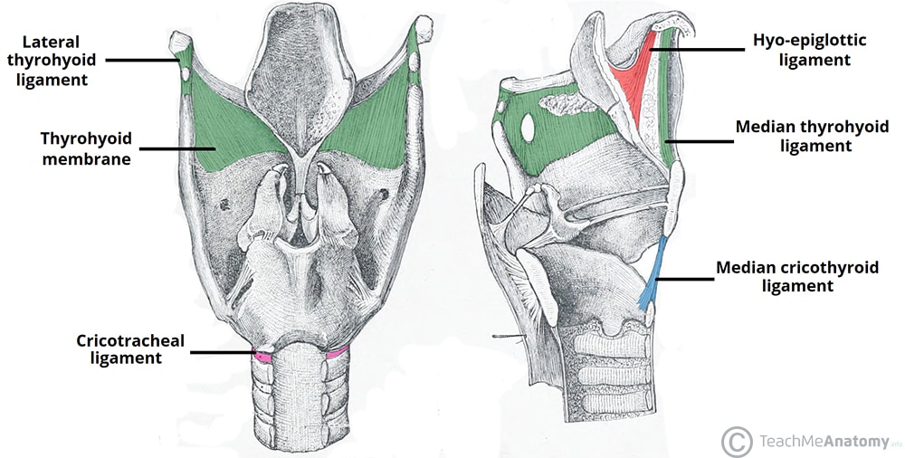
- Lateral thyrohyoid ligaments – Posterolateral thickenings of the membrane.
- Hyo-epiglottic ligament – Connects the hyoid bone to the anterior aspect of the epiglottis.
- Cricotracheal ligament – Connects the cricoid cartilage to the trachea.
- Median cricothyroid ligament – Anteromedial thickening of the cricothyroid ligament (see below), connecting the thyroid and cricoid cartilages.
Intrinsic:
- Cricothyroid ligament – Originates from the cricoid cartilage and extends superiorly, where it terminates with an free (unattached) upper margin – which forms the vocal ligament. It is additionally attached anteriorly to the thyroid cartilage, and posteriorly to the arytenoid cartilage.
- Quadrangular membrane – Spans between the anterolateral arytenoid cartilage and the lateral aspect of the epiglottis. It has a free upper margin and lower margin. The lower margin is thickened to become the vestibular ligament.
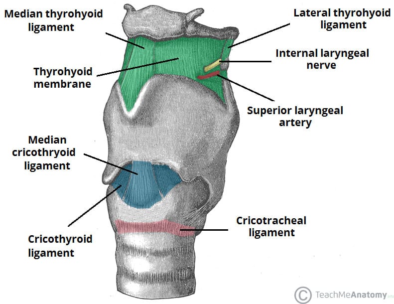
Fig 1 – Some of the major laryngeal membranes and ligaments. Note that the upper free edge of the cricothyroid ligament is not demonstrated in this image.
Clinical Relevance: Cricothyroidotomy
A cricothyroidotomy is an emergency procedure to provide a temporary airway. It is typically used in situations where there is an obstruction at or above the larynx (e.g foreign body, angioedema or facial trauma), and intubation has been unsuccessful.
To perform the technique, the thyroid cartilage is palpated in the neck – below which there is a depression representing the cricothyroid ligament. A small incision is made in the midline of this ligament, and an endotracheal tube is inserted to secure the airway.
Laryngeal Folds
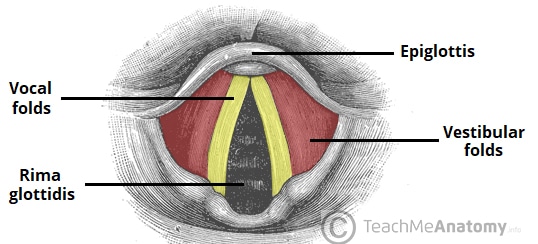
There are two important soft tissue folds located within the larynx – the vestibular folds and vocal folds. They play a crucial role in protection of the airway, breathing, and phonation.
Vocal Folds
The vocal folds (true vocal cords) are the more important of the two sets. Under the control of the muscles of phonation, they are abducted, adducted, relaxed and tensed to control the pitch of the sound created.
Histologically, they are structured as follows (superficial to deep):
- Non-keratinised stratified squamous epithelium – Stratified layer provides extensive protection against foreign bodies which may accidentally enter the larynx.
- Reinke’s space – This watery, amorphous layer is rich in glycosaminoglycans. Due to its fluidity, the epithelium is able to vibrate freely above it to create sound.
- Vocal ligament – Lies at the free upper edge of the cricothryoid ligament.
- Vocalis muscle – Exceptionally fine muscle fibres that lie lateral to the vocal ligaments.
The vocal folds are relatively avascular, and appear white in colour. The space between the vocal folds is known as the rima glottidis.
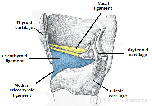
Fig 3 – The vocal ligament forms from the free upper edge of the cricothyroid ligament.
Vestibular Folds
The vestibular folds (false vocal cords) lie superiorly to the true vocal cords. They consist of the vestibular ligament (free lower edge of the quadrangular membrane) covered by a mucous membrane, and are pink in colour. They are fixed folds, which act to provide protection to the larynx.