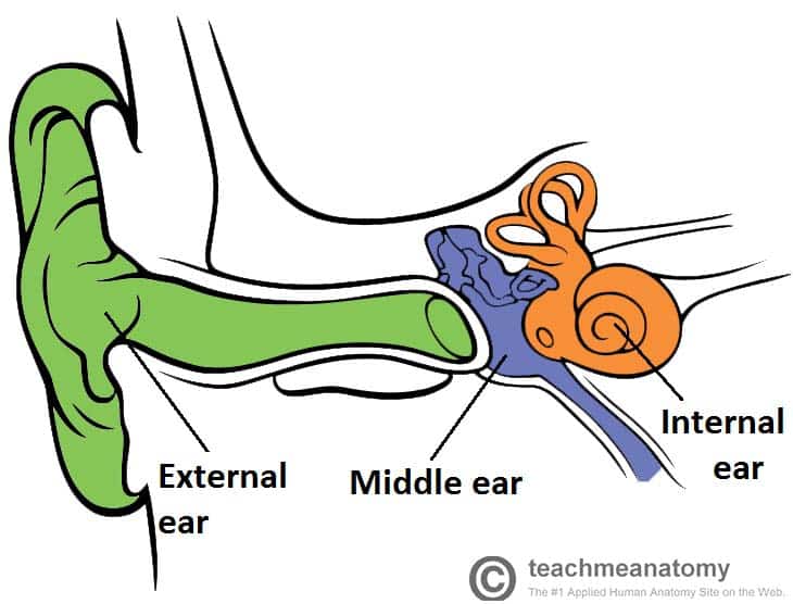
The ear can be split into three parts; external, middle and inner.
The middle ear lies within the temporal bone, and extends from the tympanic membrane to the lateral wall of the inner ear. The main function of the middle ear is to transmit vibrations from the tympanic membrane to the inner ear via the auditory ossicles.
This article will focus on the anatomy of the middle ear – its structure, neurovasculature, and its clinical correlations.
Parts of the Middle Ear
The middle ear can be divided into two parts:
- Tympanic cavity – located medially to the tympanic membrane. It contains three small bones known as the auditory ossicles: the malleus, incus and stapes. They transmit sound vibrations through the middle ear.
- Epitympanic recess – a space superior to the tympanic cavity, which lies next to the mastoid air cells. The malleus and incus partially extend upwards into the epitympanic recess.
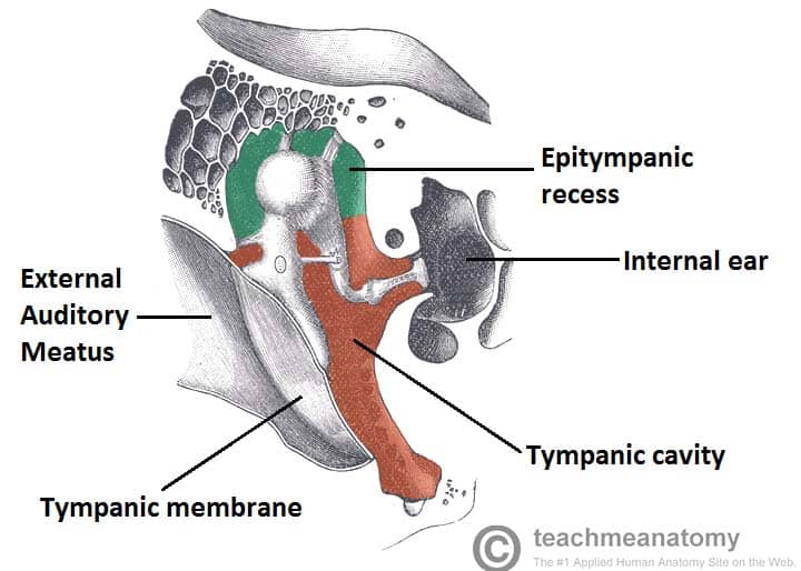
Fig 2 – The middle ear. The two main parts of the middle ear have been labelled.
Borders
The middle ear can be visualised as a rectangular box, with a roof and floor, medial and lateral walls and anterior and posterior walls.
- Roof – formed by a thin bone from the petrous part of the temporal bone. It separates the middle ear from the middle cranial fossa.
- Floor – known as the jugular wall, it consists of a thin layer of bone, which separates the middle ear from the internal jugular vein
- Lateral wall – made up of the tympanic membrane and the lateral wall of the epitympanic recess.
- Medial wall – formed by the lateral wall of the internal ear. It contains a prominent bulge, produced by the facial nerve as it travels nearby.
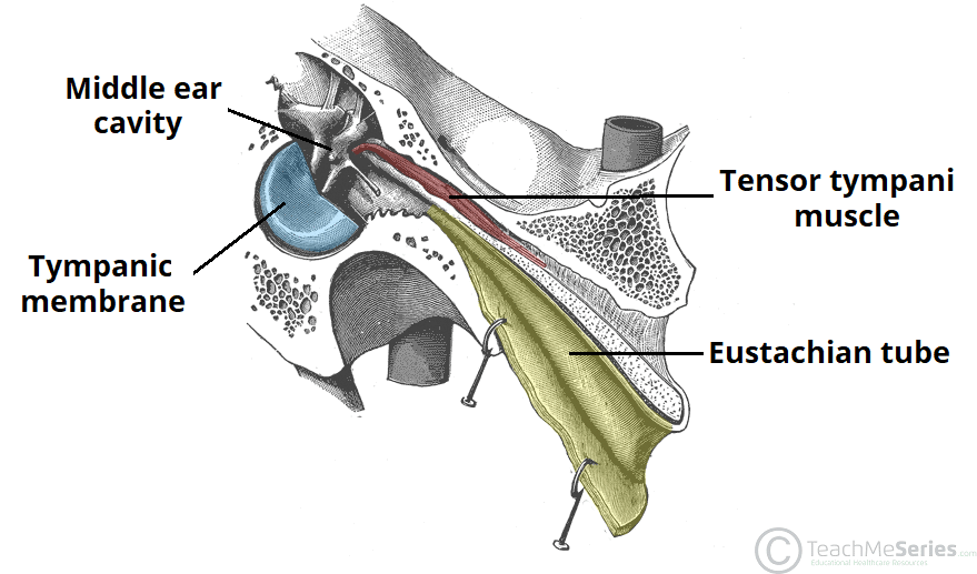
- Anterior wall – a thin bony plate with two openings; for the auditory tube and the tensor tympani muscle. It separates the middle ear from the internal carotid artery.
- Posterior wall (mastoid wall) – it consists of a bony partition between the tympanic cavity and the mastoid air cells.
- Superiorly, there is a hole in this partition, allowing the two areas to communicate. This hole is known as the aditus to the mastoid antrum.
Bones
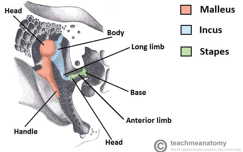
The bones of the middle ear are the auditory ossicles – the malleus, incus and stapes. They are connected in a chain-like manner, linking the tympanic membrane to the oval window of the internal ear.
Sound vibrations cause a movement in the tympanic membrane which then creates movement, or oscillation, in the auditory ossicles. This movement helps to transmit the sound waves from the tympanic membrane of external ear to the oval window of the internal ear.
The malleus is the largest and most lateral of the ear bones, attaching to the tympanic membrane, via the handle of malleus. The head of the malleus lies in the epitympanic recess, where it articulates with the next auditory ossicle, the incus.
The next bone – the incus – consists of a body and two limbs. The body articulates with the malleus, the short limb attaches to the posterior wall of the middle, and the long limb joins the last of the ossicles; the stapes.
The stapes is the smallest bone in the human body. It joins the incus to the oval window of the inner ear. It is stirrup-shaped, with a head, two limbs, and a base. The head articulates with the incus, and the base joins the oval window.
Mastoid Air Cells
The mastoid air cells are located posterior to epitympanic recess. They are a collection of air-filled spaces in the mastoid process of the temporal bone. The air cells are contained within a cavity called the mastoid antrum. The mastoid antrum communicates with the middle ear via the aditus to mastoid antrum.
The mastoid air cells act as a ‘buffer system‘ of air – releasing air into the tympanic cavity when the pressure is too low.
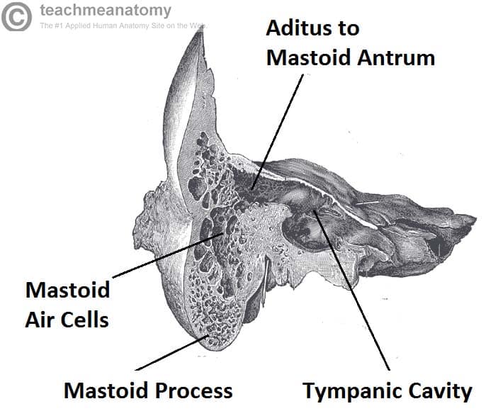
Fig 4 – Coronal section of temporal bone, showing the mastoid air cells in more detail
Muscles
There are two muscles which serve a protective function in the middle ear; the tensor tympani and stapedius. They contract in response to loud noise, inhibiting the vibrations of the malleus, incus and stapes, and reducing the transmission of sound to the inner ear. This action is known as the acoustic reflex.
The tensor tympani originates from the auditory tube and attaches to the handle of malleus, pulling it medially when contracting. It is innervated by the tensor tympani nerve, a branch of the mandibular nerve. The stapedius muscle attaches to the stapes, and is innervated by the facial nerve.
Auditory Tube
The auditory tube (eustachian tube) is a cartilaginous and bony tube that connects the middle ear to the nasopharynx. It acts to equalise the pressure of the middle ear to that of the external auditory meatus.
It extends from the anterior wall of the middle ear, in an anterior, medioinferior direction, opening onto the lateral wall of the nasopharynx. In joining the two structures, it is a pathway by which an upper respiratory infection can spread into the middle ear.
The tube is shorter and straighter in children, therefore middle ear infections tend to be more common in children than adults.
Clinical Relevance: Otitis Media with Effusion
Otitis media with effusion is commonly known as glue ear. It arises from persistent dysfunction of the auditory tube. If the auditory tube is unable to equalise middle ear pressure (due to blockage, inflammation, genetic mutation), a negative pressure develops inside the middle ear
This negative pressure draws out a transudate from the mucosa of the middle ear, creating an environment suitable for pathogens to replicate and cause infection.
Upon inspection of a patient with otitis media with effusion, the eardrum will appear inverted, with fluid visible inside the ear.
Clinical Relevance: Mastoiditis
Middle ear infections (otitis media) can spread to the mastoid air cells. Due to their porous nature, they are a suitable site for pathogenic replication.
The mastoid process itself can get infected, and this can spread to the middle cranial fossa, and into the brain, causing meningitis.
If mastoiditis is suspected, the pus must be drained from the air cells. When doing so, care must be taken not to damage the nearby facial nerve.
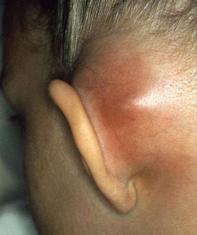
Fig 6 – Mastoiditis